Fibroblast Cells: Confocal Microscopy Digital Image Gallery
Grey Fox Lung Fibroblast Cells (FoLu Line)
The FoLu line of cells was established from the lung tissue of an adult female grey fox (Urocyon cinereoargenteus). The cells exhibit many of the typical characteristics associated with fibroblasts and are primarily used in virus studies. Many lung-derived cell lines similar to FoLu have also been utilized in studies focusing upon the short- and long-term effects of cigarette smoke, and this particular line is potentially useful for such research as well.
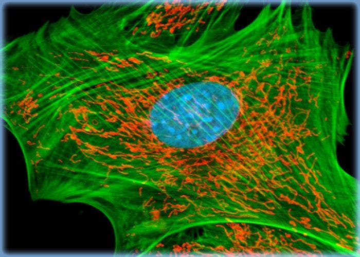
F-Actin, Mitochondria, and DNA Distribution in FoLu Fibroblasts
A log phase culture of fox lung fibroblast cells was labeled for F-actin and the intracellular mitochondrial network with Alexa Fluor 488 conjugated to phalloidin and MitoTracker Red CMXRos, respectively. DRAQ5 (pseudocolored cyan), a far-red fluorescent DNA probe, was employed as a nuclear counterstain.
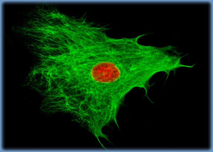
Intermediate Filaments in FoLu Fox Lung Fibroblasts
In order to label the intermediate filaments in the log phase adherent FoLu culture shown in this confocal image, the fixed and permeabilized cells were blocked and treated with mouse anti-vimentin (porcine eye lens) primary antibodies followed by goat anti-mouse secondary antibodies (IgG) conjugated to Alexa Fluor 488. Cell nuclei were counterstained with the red-absorbing dye TO-PRO-3.
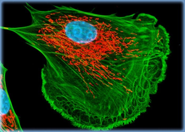
FoLu Cells Labeled for F-Actin with an Alexa Fluor Probe Conjugated to a Phallotoxin
The culture of FoLu fibroblasts presented in this section was labeled for mitochondria with MitoTracker Red CMXRos and for the cytoskeletal filamentous actin network with Alexa Fluor 488 conjugated to phalloidin, a cyclic peptide derived from the toxic death cap mushroom (Amanita phalloides). In addition, the cells were probed for DNA in the cell nucleus with TO-PRO-3, a monomeric cyanine nucleic acid stain.
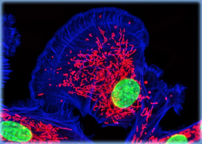
Fox Lung Fibroblast Cells (FoLu Line)
Established from the lung tissue of an adult female grey fox (Urocyon cinereoargenteus), FoLu cells exhibit many of the typical characteristics associated with fibroblasts and are primarily used in virus studies. Many lung-derived cell lines similar to FoLu have also been utilized in studies focusing upon the short- and long-term effects of cigarette smoke, and this particular line is potentially useful for such research as well.
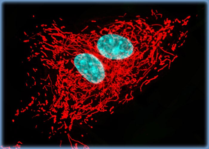
Visualizing the Proximity Between the Nucleus and the Mitochondrial Network in Fox Lung Cell Cultures
Details of the mitochondrial network in fox lung fibroblast cells were revealed by treating a FoLu culture with MitoTracker Red CMXRos, a popular red-fluorescent mitochondrion-selective stain. The high-affinity DNA probe DRAQ5 (pseudocolored cyan) was utilized to counterstain cell nuclei in the culture.
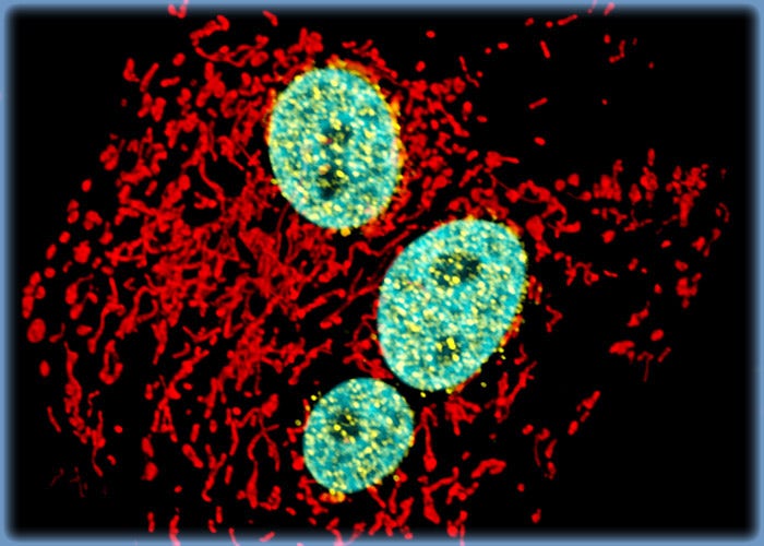
Targeting Nuclear Pore Complexes in Fox Lung Fibroblast Cells with Immunofluorescence
Nuclei in a culture of FoLu cells were stained with Hoechst 33342 and immunofluorescence was utilized to visualize the numerous nuclear pore complexes located on each nucleus. The cells were fixed, permeabilized, blocked with 10-percent normal goat serum, and treated with mouse anti-NPCP (nuclear pore complex protein) primary antibodies followed by goat anti-mouse secondary antibodies (IgG) conjugated to Alexa Fluor 647 (pseudocolored yellow). The mitochondrial network was also targeted by treating the fibroblasts with MitoTracker Red CMXRos.
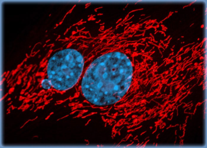
Details of the Mitochondrial Network in Fox Lung Cells Revealed with a MitoTracker Probe
MitoTracker Red CMXRos is a derivative of X-rosamine that selectively binds with mitochondria. By treating a culture of fox lung cells with the MitoTracker probe, details of the mitochondrial network were visualized. The fibroblasts were also stained with DRAQ5 (pseudocolored cyan), a far-red fluorescent DNA probe, in order to image cell nuclei.
Mongolian Gerbil Lung Fibroblast Cells (GeLu Line)
Lung tissue of a rodent native to parts of Africa and Asia, the Mongolian gerbil (Meriones unguiculatus), served as the source of the initial cells from which the GeLu fibroblast line was established. The tissue sample was excised from a 403-day-old female.
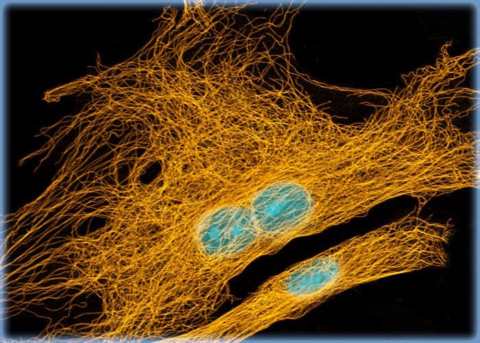
Mongolian Gerbil Lung Fibroblast Cells (GeLu Line)
Lung tissue of a rodent native to parts of Africa and Asia, the Mongolian gerbil (Meriones unguiculatus), served as the source of the initial cells from which the GeLu fibroblast line was established. The tissue sample was excised from a 403-day-old female.
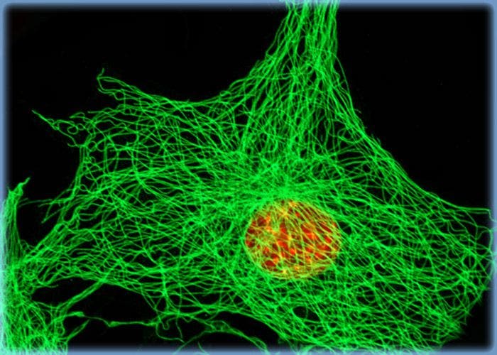
The Microtubule Network in Gerbil Lung Cells
Immunofluorescence with mouse anti-alpha-tubulin was employed to visualize details of the microtubule network in the log phase monolayer culture of gerbil lung cells presented in this section. The secondary antibody (goat anti-mouse IgG) was conjugated to Alexa Fluor 555 (pseudocolored green). Nuclei were counterstained with TO-PRO-3, a carbocyanine monomer with long-wavelength red fluorescence.
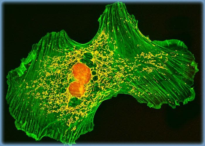
Visualizing the Actin Cytoskeleton in Gerbil Lung Cells with BODIPY FL Conjugated to a Phallotoxin
Filamentous actin was targeted in a culture of GeLu fibroblasts with BODIPY FL conjugated to phallacidin, a bicyclic peptide derived from a toxic mushroom. In addition, the cells were treated with the mitochondrion-selective stain MitoTracker Red CMXRos and a nuclear counterstain, TO-PRO-3.
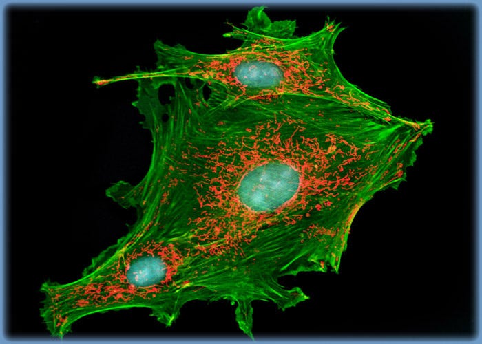
GeLu Cells with MitoTracker Red CMXRos, BODIPY FL, and TO-PRO-3
A triple fluorophore combination of MitoTracker Red CMXRos, BODIPY FL conjugated to phallacidin, and TO-PRO-3 was used to label an adherent log phase culture of Mongolian gerbil lung cells for mitochondria, the F-actin network, and nuclear DNA, respectively. The cells were first treated with the MitoTracker probe in growth medium for one hour, washed and fixed with paraformaldehyde, permeabilized, and blocked with bovine serum albumen. The cells were subsequently labeled with the conjugated phallacidin and counterstained with the cyanine monomer (TO-PRO-3) reagent.
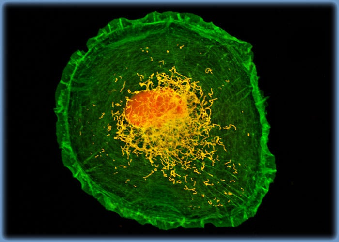
Distribution of Filamentous Actin, Mitochondria, and DNA in GeLu Fibroblasts
An adherent monolayer GeLu cell culture was labeled for cytoskeletal filamentous actin and mitochondria with Alexa Fluor 488 conjugated to phalloidin and MitoTracker Red CMXRos, respectively. Nuclei present in the fibroblast cells were counterstained with the red-absorbing DNA probe DRAQ5.
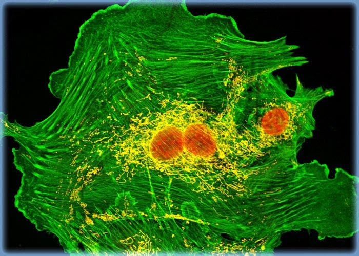
Targeting the F-Actin Cytoskeleton in GeLu Cells with a Fluorescent Probe Conjugated to a Phallotoxin
The proximity between the F-actin and mitochondrial networks was revealed in Mongolian gerbil lung cells by staining a GeLu culture with Alexa Fluor 488 conjugated to a phallotoxin (phalloidin) and MitoTracker Red CMXRos (pseudocolored yellow). The red-absorbing dye TO-PRO-3 was used to counterstain the cells for DNA in the nucleus.
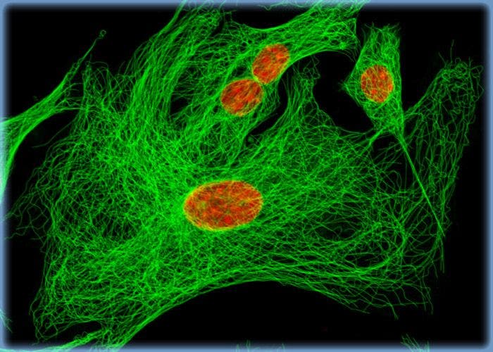
Microtubules Immunofluorescently Visualized in Mongolian Gerbil Lung Fibroblasts
The gerbil lung cells presented in this confocal image were resident in a culture fixed with 3.7 percent paraformaldehyde, permeabilized with Triton X-100, blocked with 10 percent normal goat serum and treated with mouse anti-alpha-tubulin primary antibodies. Goat anti-mouse secondary antibodies conjugated to Alexa Fluor 568 (pseudocolored green) were mixed with the DNA stain DRAQ5 in order to simultaneously visualize microtubules and cell nuclei.
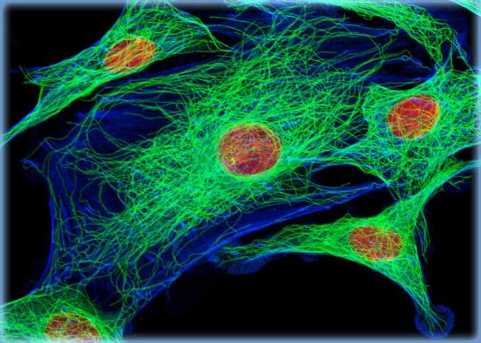
Gerbil Lung Cells Labeled for Tubulin with Immunofluorescence
In order to visualize the microtubules present in the gerbil lung cells shown in this confocal image, a GeLu culture was immunofluorescently labeled with anti-tubulin mouse monoclonal primary antibodies followed by goat anti-mouse Fab fragments conjugated to Alexa Fluor 488. In addition, the cells were labeled for the cytoskeletal F-actin network with Alexa Fluor 546 (pseudocolored blue) conjugated to phalloidin, and for cell nuclei with TO-PRO-3.
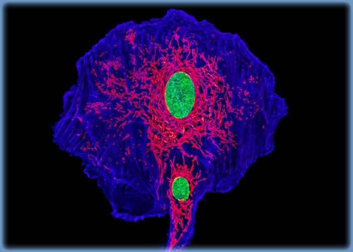
GeLu Fibroblasts Triple Labeled with MitoTracker Deep Red 633, Alexa Fluor 568, and YO-PRO-1
The Mongolian gerbil lung cells appearing in this confocal image were resident in a culture labeled with Alexa Fluor 568 (pseudocolored blue) conjugated to phalloidin, targeting the F-actin cytoskeletal network, and MitoTracker Deep Red 633, which preferentially binds with intracellular mitochondria. DNA in the nucleus was counterstained with YO-PRO-1.
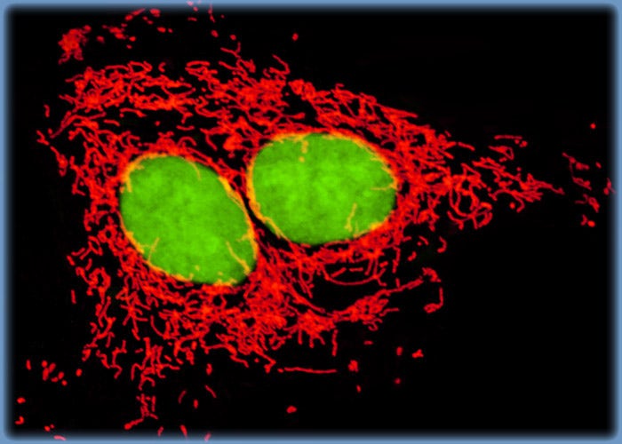
Revealing Details of the Mitochondrial Network in Mongolian Gerbil Lung Cell Cultures with MitoTracker Orange CMTMRos
The culture of GeLu fibroblast cells shown in this confocal image was labeled for the mitochondrial network with MitoTracker Orange CMTMRos (pseudocolored red), a derivative of tetramethylrosamine. The cells were also treated with the green-fluorescent nuclear and chromosome counterstain SYTOX Green.
Raccoon Uterus Fibroblast Cells (Pl 1 UT Line)
Studies have shown that Pl 1 UT cells are susceptible to an array of viruses, including herpes simplex virus, reovirus 3, and vesicular stomatitis (Ogden strain). The line is, therefore, commonly utilized in the propagation of such viruses for research purposes and has been particularly useful in investigations of feline and canine viral diseases.
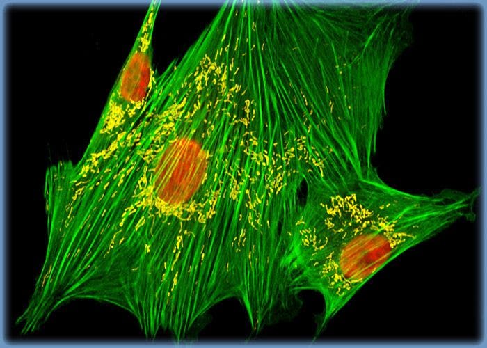
Raccoon Uterus Fibroblast Cells (Pl 1 UT Line)
The Pl 1 UT cell line was derived from the uterine tissue of an adult female North American raccoon (Procyon lotor). The cells exhibit fibroblast-like morphological characteristics and grow adherently to glass and polymer surfaces in culture.
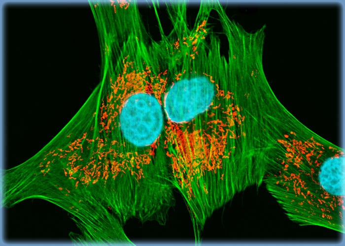
Targeting F-Actin and Mitochondria in Pl 1 UT Cell Cultures
A triple fluorophore combination of MitoTracker Red CMXRos, BODIPY FL conjugated to phallacidin, and DRAQ5 (pseudocolored cyan) was used to label an adherent log phase culture of raccoon uterus cells for mitochondria, the filamentous actin network, and nuclear DNA, respectively. The cells were first treated with the MitoTracker probe in growth medium for one hour, washed and fixed with paraformaldehyde (prepared in growth medium), permeabilized, and blocked with bovine serum albumen. The cells were subsequently labeled with the conjugated phallotoxin and counterstained with the anthraquinone (DRAQ5) reagent.
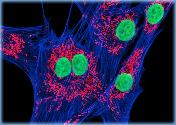
Raccoon Uterus Fibroblast Cells with BODIPY FL, MitoTracker Orange CMTMRos, and DRAQ5
The mitochondrial and cytoskeletal filamentous actin networks were targeted in the culture of Pl 1 UT fibroblasts presented in this section with BODIPY FL conjugated to phallacidin (a mushroom toxin) and MitoTracker Orange CMTMRos, respectively. Nuclei in the cells were counterstained with the far-red fluorescent DNA probe DRAQ5.
Swiss Mouse Embryo Moloney Murine Leukemia Virus Transfected Fibroblast Cells (CRE BAG 2 Line)
CRE BAG 2 is a fibroblast cell line that was developed from the NIH 3T3 embryonic Swiss mouse cell line, which was transfected with Moloney murine leukemia virus-derived proviral genomes carrying complementary mutations in the gag-pol or env regions. The genomes were altered at the 3’ end of the provirus and contained a deletion of the psi sequence needed for the efficient encapsidation of retroviral genomes into virions.
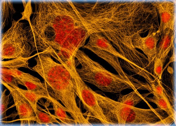
Swiss Mouse Embryo Moloney Murine Leukemia Virus Transfected Fibroblast Cells (CRE BAG 2 Line)
CRE BAG 2 is a fibroblast cell line that was developed from the NIH 3T3 embryonic Swiss mouse cell line, which was transfected with Moloney murine leukemia virus-derived proviral genomes carrying complementary mutations in the gag-pol or env regions. The genomes were altered at the 3' end of the provirus and contained a deletion of the psi sequence needed for the efficient encapsidation of retroviral genomes into virions.
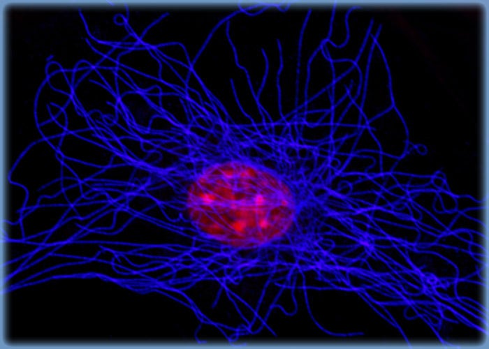
Targeting Tubulin in CRE BAG 2 Cells with Immunofluorescence
In order to visualize the microtubules present in a culture of transfected Swiss mouse fibroblasts, the cells were immunofluorescently labeled with anti-tubulin mouse monoclonal primary antibodies followed by goat anti-mouse Fab fragments conjugated to Alexa Fluor 555 (pseudocolored blue). In addition, the cells were labeled for DNA in the nucleus with the red-absorbing dye TO-PRO-3.
Embryonic Albino Swiss Mouse Fibroblast Cells (3T3 Line)
Established by George Todaro and Howard Green in 1962 from disaggregated Swiss mouse (Mus musculus) embryo tissue, the 3T3 cell line is a standard fibroblast cell line used in a wide spectrum of research and industrial biomedical applications. Variants of the initial cell line have been tested and found negative for ectromelia virus (mousepox), but most are susceptible to polyoma and simian virus 40 (SV40). In addition, 3T3 cells are negative for the viral enzyme reverse transcriptase, indicating the lack of integral retrovirus genomes.sidation of retroviral genomes into virions.
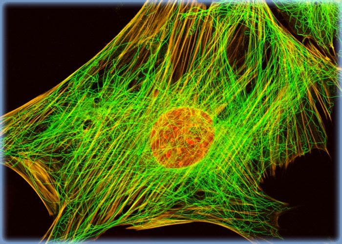
Embryonic Albino Swiss Mouse Fibroblast Cells (3T3 Line)
Established by George Todaro and Howard Green in 1962 from disaggregated Swiss mouse (Mus musculus) embryo tissue, the 3T3 cell line is a standard fibroblast cell line used in a wide spectrum of research and industrial biomedical applications. Variants of the initial cell line have been tested and found negative for ectromelia virus (mousepox), but most are susceptible to polyoma and simian virus 40 (SV40).
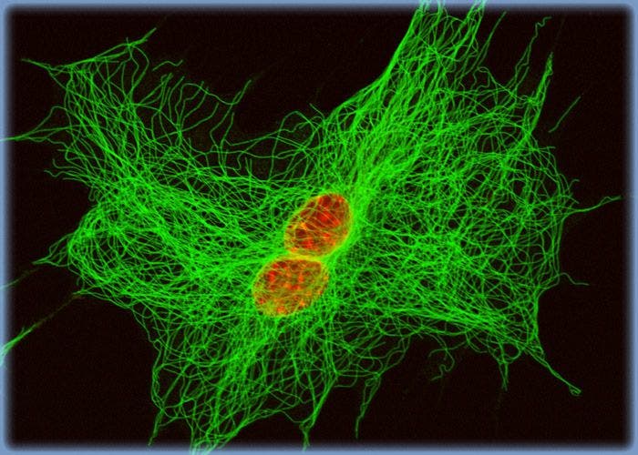
Albino Swiss Mouse Embryo Fibroblast Cells with Alexa Fluor 488 and the DNA Probe TO-PRO-3
A log phase culture of 3T3 cells was immunofluorescently labeled with primary anti-tubulin mouse monoclonal antibodies followed by goat anti-mouse secondary antibodies conjugated to Alexa Fluor 488. The cells were simultaneously stained for DNA with TO-PRO-3 and imaged using argon-ion and helium-neon lasers.
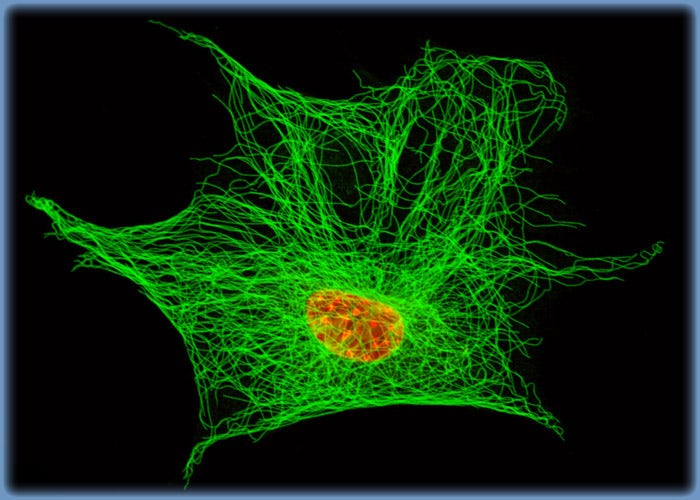
3T3 Fibroblast Cells with Alexa Fluor 488 and TO-PRO-3
Similar to one of the gallery entries listed above, an adherent log phase culture of 3T3 cells was immunofluorescently labeled with primary anti-tubulin mouse monoclonal antibodies followed by goat anti-mouse secondary antibodies conjugated to Alexa Fluor 488. The cells were simultaneously stained for DNA with TO-PRO-3 and imaged using argon-ion and helium-neon lasers.
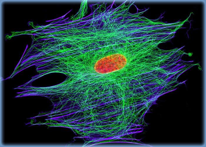
Tubulin, Actin, and DNA Distribution in 3T3 Cells
A culture of 3T3 cells was immunofluorescently labeled with primary anti-tubulin mouse monoclonal antibodies followed by goat anti-mouse secondary antibodies conjugated to Alexa Fluor 488. In addition, the cells were simultaneously probed for DNA with the red-absorbing probe TO-PRO-3, and for the cytoskeletal filamentous actin network with Alexa Fluor 568 conjugated to phalloidin.
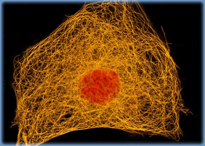
Microtubule Network in Albino Swiss Mouse Fibroblast Cells
In order to visualize the microtubules present in a culture of 3T3 fibroblasts, the cells were immunofluorescently labeled with anti-tubulin mouse monoclonal primary antibodies followed by goat anti-mouse Fab fragments conjugated to Alexa Fluor 555. In addition, the cells were labeled for DNA in the nucleus with the red-absorbing dye TO-PRO-3.
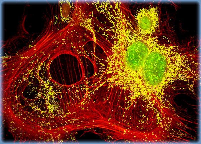
Swiss Mouse Embryo Fibroblast Cells with MitoTracker Deep Red 633, Texas Red, and SYTOX Green
A log phase culture of embryonic Swiss mouse fibroblast cells was stained with MitoTracker Deep Red 633, Texas Red conjugated to phalloidin, and SYTOX Green, which target the intracellular mitochondrial network, cytoskeletal actin filaments, and nuclei, respectively. High signal levels from all three of the fluorophores employed to stain the culture are present.
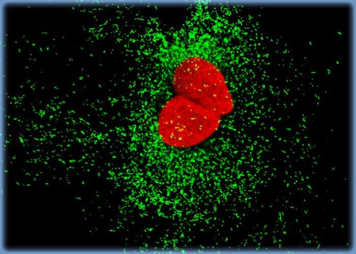
Distribution of Histone Proteins and Peroxisomes in 3T3 Cultures
Nuclear histone proteins were targeted in the culture of 3T3 cells presented in this section with mouse anti-histone (pan) monoclonal antibodies, which were imaged with goat anti-mouse Fab fragments conjugated to Alexa Fluor 568 (labeling the nucleus). The specimen was simultaneously labeled for peroxisomes with Alexa Fluor 488 conjugated to goat secondary antibodies that target rabbit anti-PMP 70 (peroxisomal membrane protein 70).
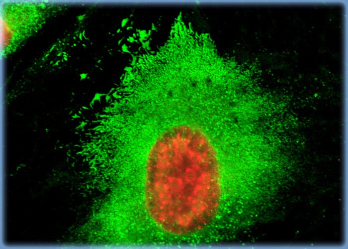
Using Concanavalin A to Probe the Endoplasmic Reticulum Network in Albino Swiss Mouse Embryo Cells
A single Swiss mouse embryo fibroblast cell resident in an adherent culture and stained with Alexa Fluor 488 conjugated to the lectin concanavalin A is presented in this section. Concanavalin A selectively binds to alpha-mannopyranosyl and alpha-glucopyranosyl residues in glycoproteins found in the endoplasmic reticulum. The specimen was also counterstained with TO-PRO-3, targeting DNA in the nuclei.
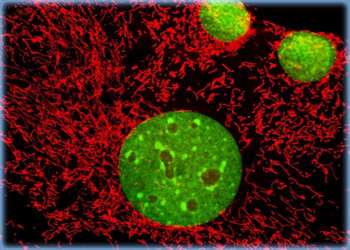
Imaging the Mitochondrial Network in Monolayer 3T3 Fibroblast Cell Cultures
A monolayer culture of Swiss mouse embryo cells was treated with MitoTracker Red CMXRos in medium containing 15 percent Cosmic calf serum, fixed with the same medium containing 3.7 percent paraformaldehyde, permeabilized with 0.2 percent Triton X-100, and then counterstained with SYTOX Green, targeting DNA in the nuclei.
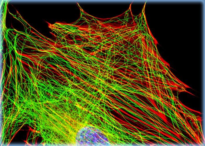
Imaging the Cytoskeletal Network in 3T3 Cell Cultures
The isolated 3T3 cell presented in this section was resident in an adherent culture stained for F-actin with Alexa Fluor 546 conjugated to phalloidin, and for DNA with the red-absorbing dye TO-PRO-3. In addition, the culture was immunofluorescently labeled with Alexa Fluor 488 conjugated to antibodies that target alpha-tubulin, a major component of the microtubule network.
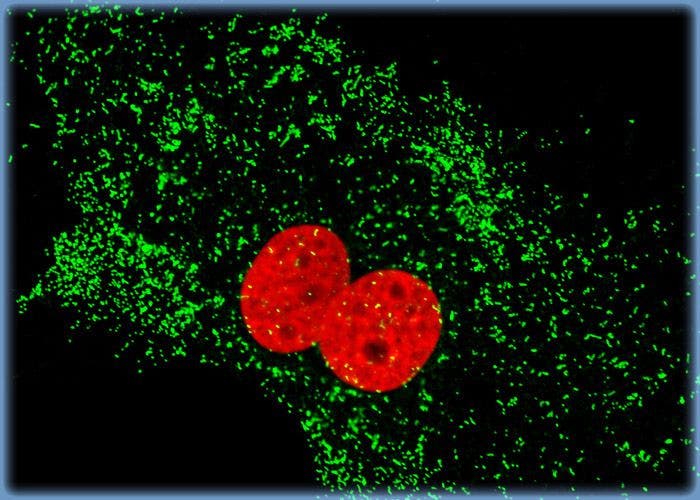
Targeting the Nuclear Histones and Cytoplasmic Peroxisomes in Albino Swiss Mouse Fibroblast Cells
In a double immunofluorescence labeling experiment, an adherent culture of Swiss mouse embryo cells was treated with a cocktail of mouse anti-histones (pan) and rabbit anti-PMP 70 (peroxisomal membrane protein) primary antibodies, followed by goat anti-mouse and anti-rabbit secondary antibodies conjugated to Alexa Fluor 568 and Alexa Fluor 488, respectively, to target the nuclear histone proteins and peroxisomes.
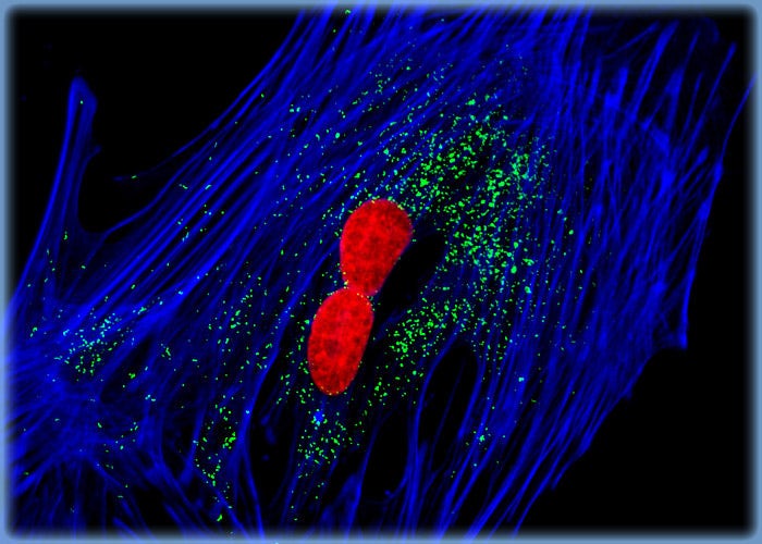
Targeting Filamentous Actin in Mouse Fibroblasts with a Phallotoxin
A log phase culture of 3T3 cells was treated with a cocktail of mouse anti-histones (pan) and rabbit anti-PMP 70 (peroxisomal membrane protein) primary antibodies, followed by goat anti-mouse and anti-rabbit secondary antibodies conjugated to Alexa Fluor 568 and Alexa Fluor 488, respectively, to target the nuclear histone proteins and peroxisomes. In addition, the F-actin network was labeled with Alexa Fluor 633 (pseudocolored blue) conjugated to phalloidin, a phallotoxin isolated from the toxic death cap mushroom.
Horse Dermal Fibroblast Cells (NBL-6 Line)
Initiated from the dermal tissue of a 4-year-old female horse (Equus caballus) of the quarterhorse strain, the NBL-6 cell line (also called the E. Derm line) has played an important role in equine viral arteritis (EVA) research. EVA is a contagious respiratory disease caused by a small, enveloped RNA virus. The fibroblast cell line is also commonly utilized to propagate viruses for equine vaccine production.
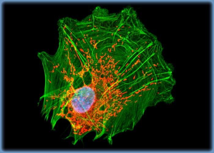
Distribution of F-Actin and Mitochondria in NBL-6 Cells
A triple fluorophore combination of MitoTracker Red CMXRos, Alexa Fluor 488 conjugated to phalloidin, and DRAQ 5 (pseudocolored cyan) was used to label an adherent log phase culture of NBL-6 cells for mitochondria, the filamentous actin network, and nuclear DNA, respectively. The cells were first treated with the MitoTracker probe in growth medium for one hour, washed and fixed with paraformaldehyde (prepared in growth medium), permeabilized, and blocked with bovine serum albumen.
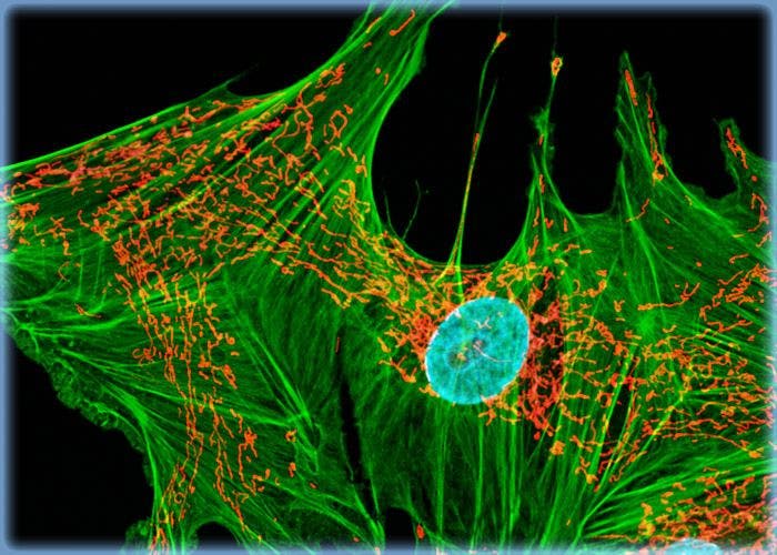
Horse Dermal Fibroblast Cells (NBL-6 Line)
Initiated from the dermal tissue of a 4-year-old female horse (Equus caballus) of the quarterhorse strain, the NBL-6 cell line (also called the E. Derm line) has played an important role in equine viral arteritis (EVA) research. EVA is a contagious respiratory disease caused by a small, enveloped RNA virus.
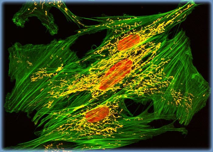
Horse Dermal Fibroblasts Triple Labeled with BODIPY FL, MitoTracker Orange CMTMRos, and TO-PRO-3
The distribution of filamentous actin and mitochondria in a culture of horse dermal fibroblast cells was visualized with BODIPY FL conjugated to phallacidin and MitoTracker Orange CMTMRos (pseudocolored yellow). Cell nuclei were counterstained with TO-PRO-3, a carbocyanine monomer with long-wavelength red fluorescence.
Human Fetal Lung Fibroblast Cells (MRC-5 Line)
MRC-5 cells are a line of human lung fibroblast cells established in the 1960s by J. P. Jacobs. Studies have demonstrated that the cells are susceptible to several different viruses, including vesicular stomatitis (Indiana strain), poliovirus 1, and herpes simplex. The MRC-5 line is frequently used in laboratories for many different applications, such as in vitro cytotoxicity testing, the development of vaccines, and as a transfection host for investigation of viruses and viral diseases.
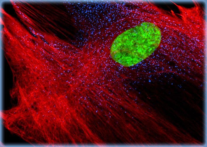
Human Fetal Lung Fibroblast Cells (MRC-5 Line)
MRC-5 cells are a line of human lung fibroblast cells established in the 1960s by J. P. Jacobs. Studies have demonstrated that the cells are susceptible to several different viruses, including vesicular stomatitis (Indiana strain), poliovirus 1, and herpes simplex.
Indian Muntjac Deer Skin Fibroblast Cells
A fibroblast cell line established from a skin biopsy of an adult male, the Indian Muntjac deer epidermis line is commonly used in laboratories around the world, especially for chromosome studies. The normal (non-transformed) Indian Muntjac cell line is susceptible to the herpes simplex virus, vaccinia virus, and vesicular stomatitis virus (Indiana strain).
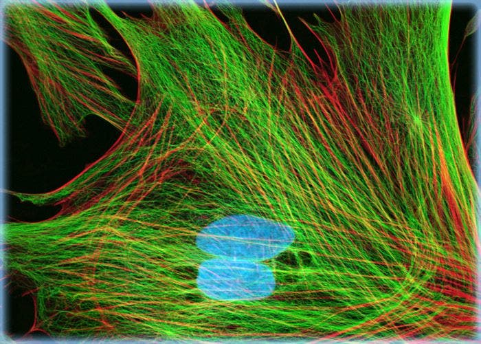
Indian Muntjac Deer Skin Fibroblast Cells
A fibroblast cell line established from a skin biopsy of an adult male, the Indian Muntjac deer epidermis line is commonly used in laboratories around the world, especially for chromosome studies. Members of the family Cervidae, Muntjacs are barking deer that emit their characteristic sound when they feel threatened or alarmed.
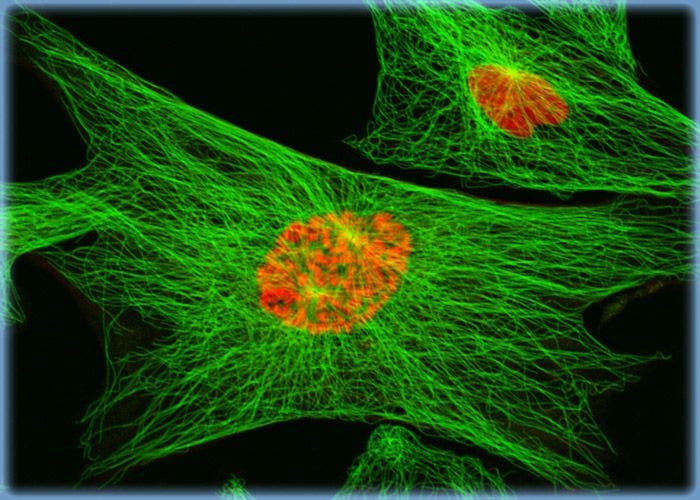
Distribution of F-Actin and Mitochondria in Muntjac Cell Cultures
The proximity between the mitochondrial network and the filamentous actin cytoskeleton was visualized by treating a culture of Indian Muntjac deer skin fibroblasts with MitoTracker Red CMXRos and Alexa Fluor 488 conjugated to phalloidin. The specimen was also probed with DRAQ5, a DNA-interactive reagent that exhibits preferential intercalation at AT base pairs.
Imaging the Mitochondria Network, Filamentous Actin, and Nuclei in Indian Muntjac Deer Skin Fibroblast Cells
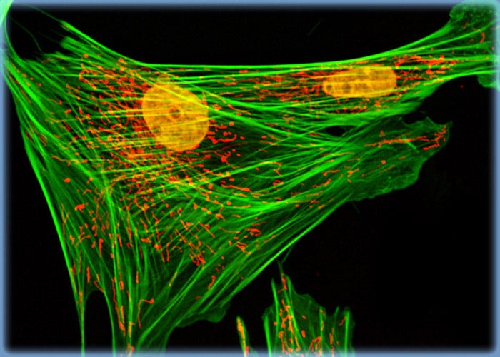
A triple fluorophore combination of MitoTracker Deep Red 633, BODIPY FL conjugated to phalloidin, and SYTOX Orange was used to label an adherent log phase culture of Indian Muntjac cells for mitochondria, the filamentous actin network, and nuclear DNA, respectively. The cells were first treated with MitoTracker Deep Red 633 in growth medium for one hour, washed and fixed with paraformaldehyde (prepared in growth medium), permeabilized, and blocked with bovine serum albumen. The cells were subsequently labeled with the conjugated phalloidin and counterstained with the SYTOX reagent.
MitoTracker, Actin, and DNA Probes with Indian Muntjac Deer Skin Fibroblast Cells
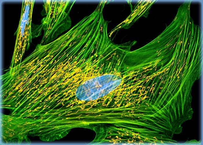
A triple fluorophore combination of MitoTracker Orange CMTMRos, BODIPY FL conjugated to phalloidin, and TO-PRO-3 was used to label an adherent log phase culture of Indian Muntjac cells for mitochondria, the filamentous actin network, and nuclear DNA, respectively. The cells were first treated with the MitoTracker probe in growth medium for one hour, washed and fixed with paraformaldehyde (prepared in growth medium), permeabilized, and blocked with bovine serum albumen. The cells were subsequently labeled with the conjugated phalloidin and counterstained with the cyanine monomer (TO-PRO-3) reagent.
The Metaphase Spindle in Dividing Indian Muntjac Cells
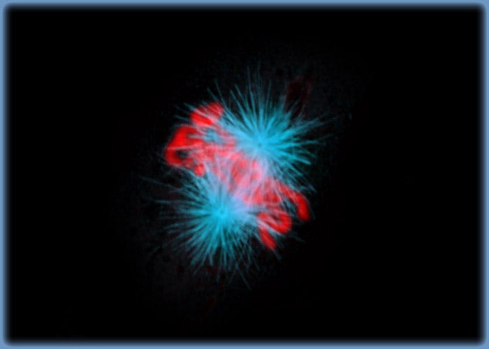
The metaphase spindle apparatus is revealed in this confocal image of Indian Muntjac deer skin cells stained with Alexa Fluor 568 for microtubules and TO-PRO-3 for DNA. The cells were fixed with 3.7 percent paraformaldehyde, permeabilized with Triton X-100, blocked with 10 percent normal goat serum and treated with mouse anti-alpha-tubulin primary antibodies. Goat anti-mouse secondary antibodies conjugated to Alexa Fluor 568 were mixed with the DNA stain.
The Mitotic Apparatus in Dividing Indian Muntjac Deer Skin Cells
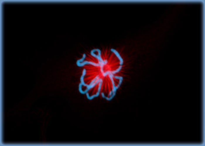
The first stages of mitosis are revealed in the confocal image of Indian Muntjac deer skin cells presented in this section, which were stained with Alexa Fluor 568 (tubulin) and TO-PRO-3 (DNA). The cells were fixed with 3.7 percent paraformaldehyde, permeabilized with Triton X-100, blocked with 10 percent normal goat serum and treated with mouse anti-alpha-tubulin primary antibodies. Goat anti-mouse secondary antibodies conjugated to Alexa Fluor 568 were mixed with the DNA stain.
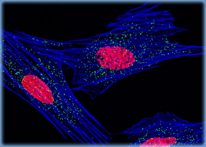
Peroxisome and Histone Protein Distribution in Muntjac Fibroblasts
In a double immunofluorescence labeling experiment, a culture of Indian Muntjac fibroblasts was treated with a cocktail of mouse anti-histones (pan) and rabbit anti-PMP 70 (peroxisomal membrane protein) primary antibodies, followed by goat anti-mouse and anti-rabbit secondary antibodies conjugated to Alexa Fluor 568 and Alexa Fluor 488, respectively, to target the nuclear histone proteins and peroxisomes. The actin cytoskeleton was visualized with Alexa Fluor 633 conjugated to phalloidin.
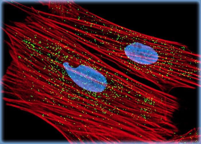
Visualizing the Nuclear Histones and Cytoplasmic Peroxisomes in Indian Muntjac Cell Cultures
A log phase culture of Indian Muntjac cells was treated with a cocktail of mouse anti-histones (pan) and rabbit anti-PMP 70 (peroxisomal membrane protein) primary antibodies, followed by goat anti-mouse and anti-rabbit secondary antibodies conjugated to Alexa Fluor 647 and Alexa Fluor 488, respectively, to target the nuclear histone proteins and peroxisomes. In addition, the F-actin network was labeled with Alexa Fluor 568 conjugated to phalloidin, a phallotoxin isolated from the death cap mushroom.
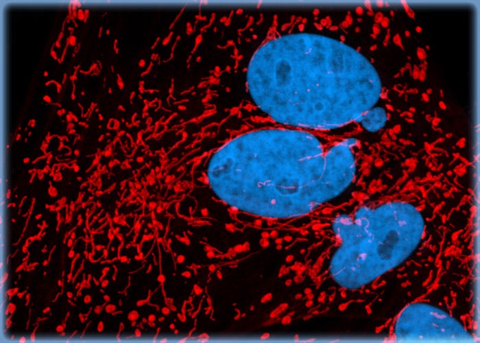
Targeting the Mitochondria Network and Nuclear DNA in Indian Muntjac Deer Skin Fibroblasts
The monolayer culture of Indian Muntjac fibroblast cells shown in this section was treated with MitoTracker Red CMXRos in medium containing 15 percent Cosmic calf serum, fixed with the same medium containing 3.7 percent paraformaldehyde, permeabilized with 0.2 percent Triton X-100, and then counterstained with DRAQ5 (pseudocolored cyan), targeting DNA in the cell nuclei.
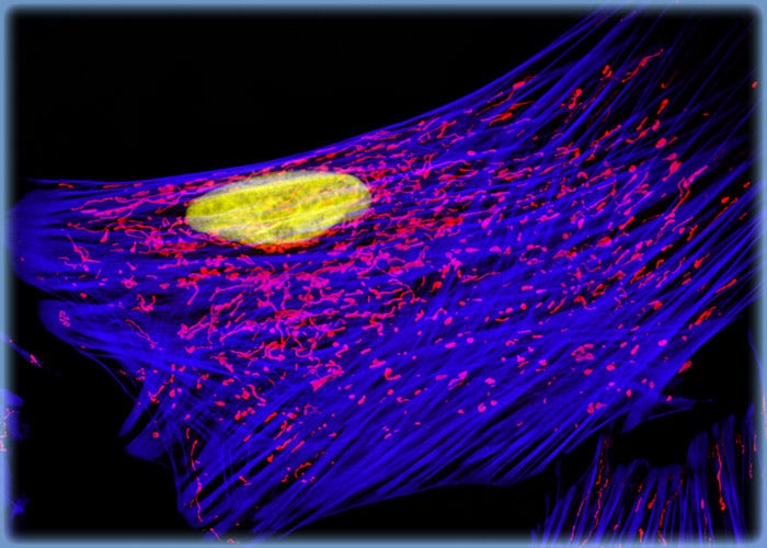
Indian Muntjac Fibroblasts with MitoTracker Red CMXRos, Alexa Fluor 488, and DRAQ5
The cytoskeletal F-actin and mitochondrial networks were targeted in the culture of Indian Muntjac cells presented in this section with Alexa Fluor 488 (pseudocolored blue) conjugated to phalloidin (a mushroom toxin) and MitoTracker Red CMXRos, respectively. Nuclei in the fibroblasts were counterstained with the far-red fluorescent DNA probe DRAQ5 (pseudocolored yellow).
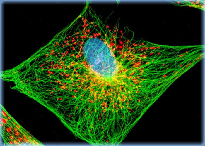
Distribution of F-Actin and Mitochondria in Muntjac Cell Cultures
The proximity between the mitochondrial network and the filamentous actin cytoskeleton was visualized by treating a culture of Indian Muntjac deer skin fibroblasts with MitoTracker Red CMXRos and Alexa Fluor 488 conjugated to phalloidin. The specimen was also probed with DRAQ5, a DNA-interactive reagent that exhibits preferential intercalation at AT base pairs.
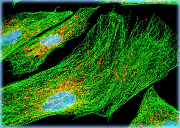
Muntjac Skin Cells Labeled for Tubulin with Immunofluorescence
In order to visualize the microtubules present in a culture of Indian Muntjac fibroblasts, the cells were immunofluorescently labeled with anti-tubulin mouse monoclonal primary antibodies followed by goat anti-mouse Fab fragments conjugated to Alexa Fluor 488. In addition, the cells were labeled for mitochondria with MitoTracker Red CMXRos, a derivative of X-rosamine, and DNA in the nucleus with the far-red fluorescent probe DRAQ5.
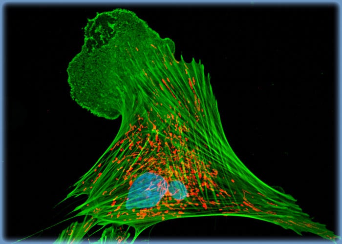
Indian Muntjac Deer Skin Cells Triple Labeled for Mitochondria, Filamentous Actin, and DNA
A triple fluorophore combination of MitoTracker Red CMXRos, Alexa Fluor 488 conjugated to phalloidin, and DRAQ5 was used to label the adherent culture of Indian Muntjac cells presented in this section for mitochondria, the F-actin network, and nuclear DNA, respectively. The cells were first treated with the MitoTracker probe in growth medium for one hour, washed and fixed with paraformaldehyde (prepared in growth medium), permeabilized, and blocked with bovine serum albumen. The cells were subsequently labeled with the conjugated phalloidin and counterstained with the anthraquinone (DRAQ5) reagent.
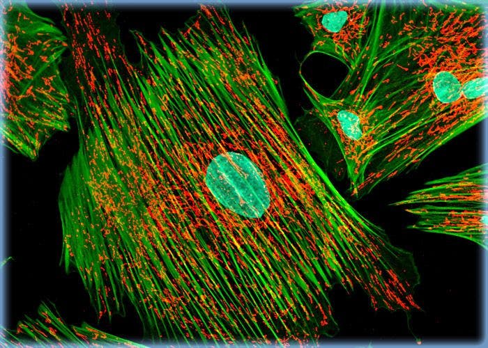
Indian Muntjac Cells with MitoTracker Red CMXRos, Alexa Fluor 488, and DRAQ 5
The adherent monolayer Indian Muntjac cell culture shown in this confocal image was labeled for the cytoskeletal filamentous actin and intracellular mitochondrial networks with Alexa Fluor 488 conjugated to phalloidin and MitoTracker Red CMXRos, respectively. Nuclei present in the fibroblasts were counterstained with the far-red fluorescent DNA-probe, DRAQ 5.
Normal African Green Monkey Kidney Fibroblast Cells (CV-1 Line)
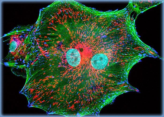
Investigators working with simian virus 40 (SV40) or in the field of AIDS research often utilize CV-1 cells, which were initially used in studies of the Rous sarcoma virus (RSV). A team led by F. C. Jensen established the CV-1 cell line in the early 1960s. The cells from which the line was initiated were sampled from the renal tissue of an African green monkey (Cercopithecus aethiops). CV-1 cells are popularly employed in transfection experiments and are known to be susceptible to a wide array of viruses.
Normal African Green Monkey Kidney Fibroblast Cells (CV-1 Line)
Investigators working with simian virus 40 (SV40) or in the field of AIDS research often utilize CV-1 cells, which were initially used in studies of the Rous sarcoma virus (RSV). A team led by F. C. Jensen established the CV-1 cell line in the early 1960s.
Transformed African Green Monkey Kidney Fibroblast Cells (COS-7 Line)
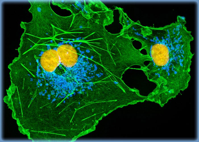
Adherent growth to both glass and plastic surfaces is characteristic of COS-7 cells in culture. The line, which is often utilized as a transfection host, retains the CV-1 trait of complete permissiveness for the lytic growth of SV40 and supports the replication of the tsA209 strain of the virus at 40 degrees Celsius as well as SV40 mutants with deletions in the early region. COS-7 cells are a popular research tool, especially for transfection experiments with recombinant plasmids.
Transformed African Green Monkey Kidney Fibroblast Cells (COS-7 Line)
The CV-1 cell line, which is permissive for lytic growth of simian virus 40 (SV40), was established in the 1960s from the kidney of an African green monkey. The COS-7 line was developed from that line about 20 years later by Yakov Gluzman via transformation with an origin defective mutant of SV40 that codes for the wild-type virus T-antigen.
Mitochondria, Actin, and DNA Distribution in COS-7 Fibroblasts
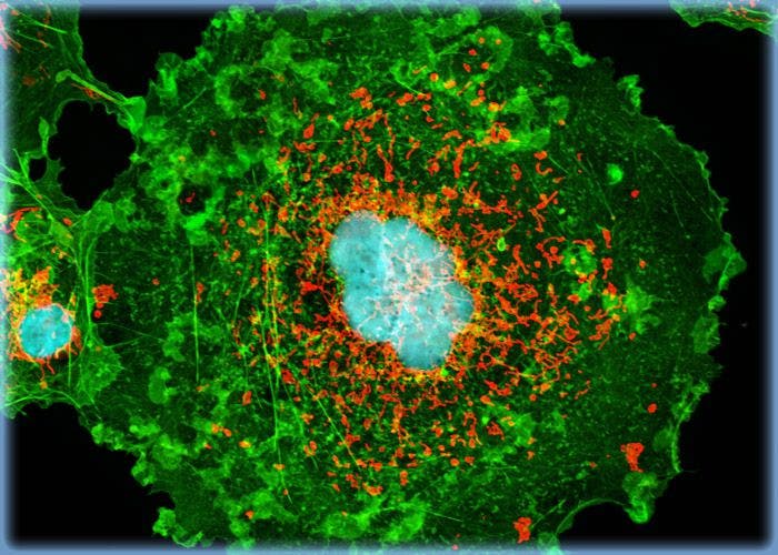
The filamentous actin and mitochondrial networks were targeted in an adherent culture of COS-7 kidney cells with BODIPY FL conjugated to phallacidin and MitoTracker Orange CMTMRos, respectively. DNA in the cells was counterstained with the red-absorbing dye TO-PRO-3 (pseudocolored cyan).
Targeting F-Actin in African Green Monkey Kidney Cell Cultures with Phallotoxins
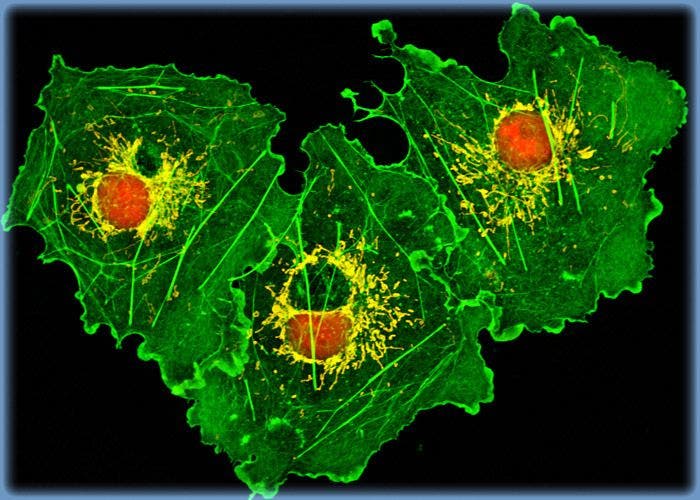
The culture of COS-7 kidney cells presented in this section was labeled for mitochondria with MitoTracker Orange CMTMRos and for the cytoskeletal filamentous actin network with BODIPY FL conjugated to phallacidin, a cyclic peptide derived from a toxic mushroom (Amanita phalloides). In addition, the cells were probed for DNA in the cell nucleus with TO-PRO-3, a monomeric cyanine nucleic acid stain.
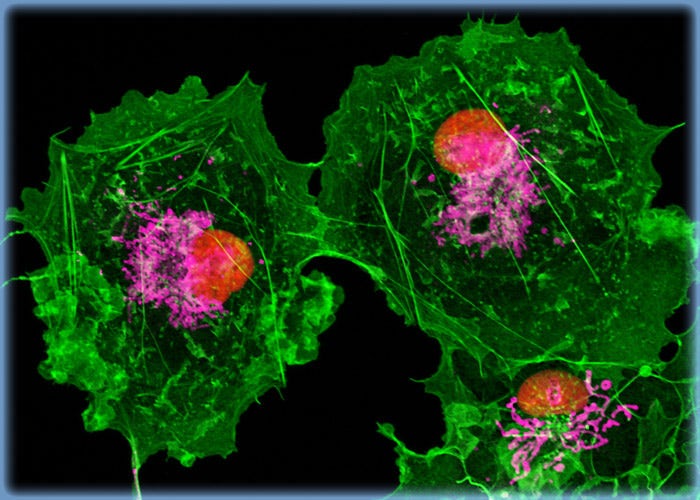
COS-7 Cells with BODIPY FL, MitoTracker Deep Red 633, and SYTOX Orange
A COS-7 fibroblast cell culture was stained with BODIPY FL conjugated to phallacidin, targeting the F-actin cytoskeletal network. The cells were also stained with MitoTracker Deep Red 633 (pseudocolored magenta) and SYTOX Orange (pseudocolored red), which preferentially bind with intracellular mitochondria and DNA in the cell nucleus, respectively.