Pramana POD Benchtop Whole Slide Scanner
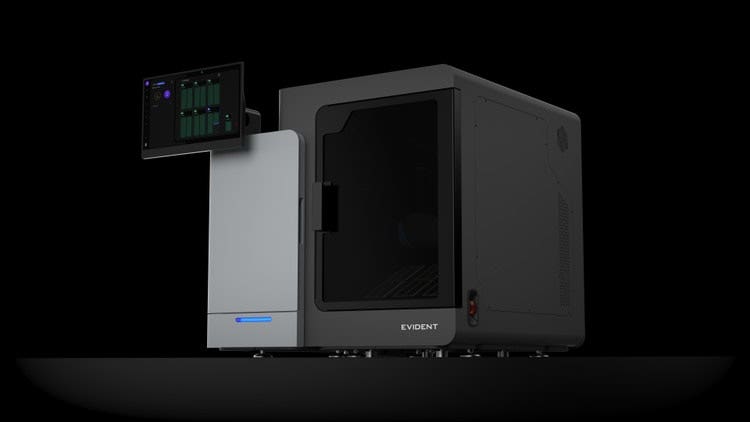
The Pramana™ POD benchtop WSI scanner helps laboratories turn most varieties of glass slides into pathologist-ready whole slide images with minimal operator intervention. Designed for small-to-medium laboratories and dedicated departments within larger organizations, the Pramana POD scanner makes digital adoption more accessible by fitting into existing workflows and lab spaces.
Pramana POD Benchtop Whole Slide Scanner
Quality Assured in Every Scan
Experience proven Pramana™ whole slide imaging technology in a smaller benchtop footprint, with automated brightfield imaging designed to support workflows for labs of varying sizes.
The Pramana POD benchtop whole slide scanner combines built-in quality assurance, true 40X imaging, and automated slide handling in one fully enclosed system.
Key Benefits of Pramana POD Scanners
Built-In Quality Assurance
In-line image-quality assessment helps identify scans that may require further review, with integrated rescan management. No separate QA software add-on or additional QA fee.
Benchtop Autonomous Imaging
Up to 300 slides in Pramana baskets or up to 200 in Sakura racks, with continuous loading and unloading options.
Built for Real-World Slides
Supports diverse specimen types and single- and double-thick slides, with acquisition that adapts to specimen characteristics.
Connected Workflows
The Pramana POD scanner is designed to operate alongside other Pramana whole slide imaging devices in multi-system environments.
Volumetric imaging in action: multiple focal planes are captured, sharp focal information is selected, and the scanned fields are stitched into a whole slide image.
Volumetric Imaging for Varied Specimens
Full-time volumetric imaging captures multiple focal planes to support the selection of sharp focal information across the scanned area. The acquisition adapts to specimen characteristics, with up to 25 quality-assured slides per hour at true 40X imaging with a 0.95 NA objective under defined conditions.
Intuitive Interface
The Pramana scanner dashboard offers an easy-to-use interface designed to streamline scanning workflows.
- Customizable settings: Adjust scanning parameters for different slide types and workflows
- Post-scan review: Review and validate scanned slides
- Native DICOM output: Supports digital laboratory workflows through native DICOM image output
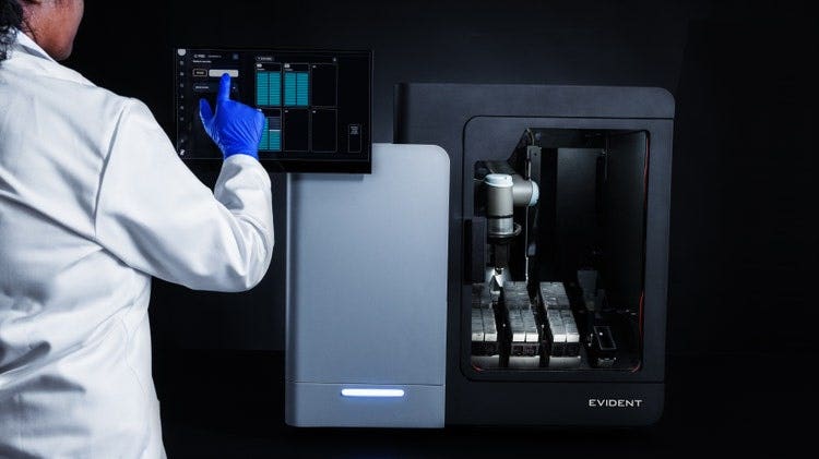
Versatility Across Pathology Applications
Supporting diverse specimen types, the Pramana POD scanner is well-suited for most brightfield imaging applications, including histopathology, hematology, cytology, and microbiology.
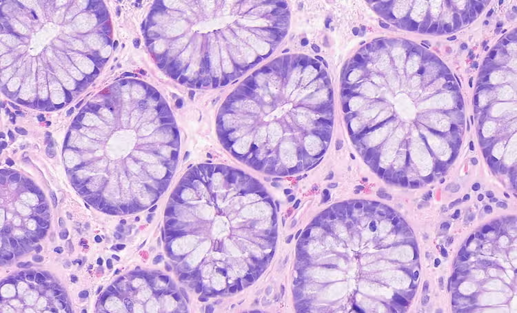
Intestine
H&E, 40X
Histopathology
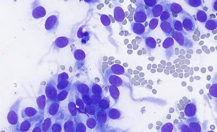
Cytology conventional smear
MGG stain, 40X
Cytology
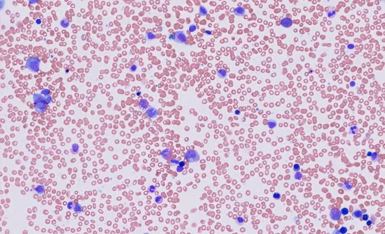
Bone marrow aspirate smear
Giemsa stain, 40X
Hematology
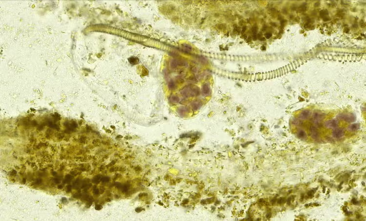
Human fecal trichrome
40X
Microbiology
Designed for Small and Medium Laboratories
The Pramana POD scanner supports laboratories introducing automated slide digitization or expanding an existing digital workflow. It provides dedicated scanning capacity for departments in larger organizations looking to manage their own slide workloads.
Expand Your Digital Pathology Capabilities
The Pramana POD scanner adds an automated benchtop option to Evident’s digital pathology portfolio. Explore our platforms designed for various slide workloads and imaging requirements to build as your needs evolve.
Specifications
0.360 micron per pixel at 20X
0.182 microns per pixel at 40X
0.120 microns per pixel at 60X
Case ID, block ID, slide name, stain name, accession date, organ / biopsy site
(L × W× H)
For Research Use Only. Not for use in diagnostic procedures. Future clinical availability is subject to applicable regulatory authorization, and regulatory status and availability may vary by country. Performance varies by specimen type and workflow.