Innovation at the Core
The System Your Users Trust
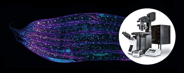
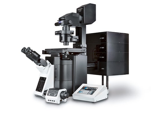
The FLUOVIEW™ FV4000 laser scanning microscope meets the rigorous demands of core imaging facilities with breakthrough technology, minimal maintenance requirements, and a user-friendly interface that makes the system fast to learn. See why the FV4000 microscope is quickly becoming the trusted choice of professionals worldwide.
FV4000 Benefits for Core Facilities
- High Performance, Low Maintenance
- Consistent Performance Every Day
- Future-Proof Your Facility with Industry-Leading Features
- Fast to Learn, Easy to Use
- Expert Support, Lasting Peace of Mind
- Precise, Reproducible Research
Trusted by Professionals
"Compared to other confocals I have experience with yours was very stress-free to use (no fear of destroying the sample or feeling overwhelmed with the software/general operation) and required a minimal amount of adjustments from the user in order to produce good quality data without feeling that my options are limited.”

Sanni Erämies | Tampere University / Imaging Facility Tampere
“The detectors are easy to set up due to their high dynamic range. They can handle low signals, but also do not switch off and perform well with high signals. This makes it very user friendly to set up the microscope. The photon counting mode is a highly appreciated function. The high precision, simple adjustment, and high flexibility make the FV4000 a very valuable tool for imaging facilities with users of different experience levels and varying complexity in their imaging experiments.”

Johannes Riemann | Center for Microscopy and Image Analysis, University of Zurich
“Together with the AI restoring tools, it gives a perfect combination for fast and high-quality volumetric imaging.”
Edwin Hernandez | Core Facility Manager, Cajal International Neuroscience Center (CINC)
“The new FV4000 will allow for faster imaging and better preservation of the sample and will allow better reproducibility.”

Alexia Ferrand | Advanced Microscopy Specialist, University of Basel
“Demonstratively better signal to noise [than other systems].”

Geoff Williams | Leduc Bioimaging Facility Manager, Brown University
“I am really impressed with the system and its performance. The super sensitivity was really impressive.”
Sara R. Roig | Advanced Microscopy Specialist, University of Basel