Exploring the Beauty of Natural Forms—6th Annual Image of the Year Americas Winner
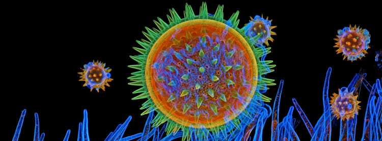
Evident Image of the Year Award 2025: Americas Winner.
Mallow pollen on stigma by Igor Siwanowicz, USA.

Igor Siwanowicz, of the United States, was named the Americas winner in Evident’s sixth Annual Image of the Year contest for his mesmerizing image of mallow pollen grains on a flower stigma.
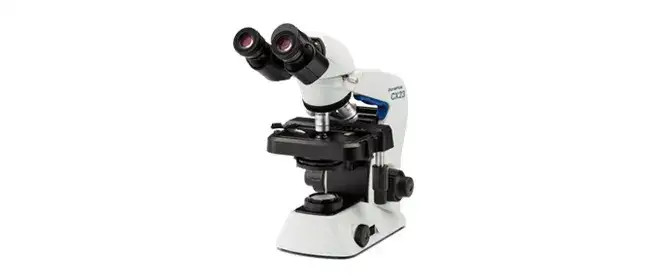
The striking specimen was collected from the flower beds at Howard Hughes Medical Institute’s Janelia Research Campus, where Igor currently specializes in invertebrate anatomy and microscopy research. The image was captured using a confocal laser scanning microscope equipped with a 40X oil immersion objective. In recognition of his winning submission, Igor will receive an Evident CX23 upright microscope.
Illuminating the Unseen Beauty of the Natural World
For Igor, beauty can be found at every scale of the natural world. Through microscopy, even a common flower can reveal extraordinary designs invisible to the naked eye. The natural forms of ordinary specimens become intricate landscapes, offering a close-up look at nature’s hidden artistry and igniting interest in both scientific experts and curious observers.
Igor, congratulations on being named the Image of the Year Americas winner! What do you find personally exciting about this image?
Natural forms, at every scale, have always attracted my attention and ignited interest. In this specific case, I was curious to see how relatively giant (~0.1 mm in diameter) mallow pollen grains germinate on the stigma. I wanted to capture the formation of the pollen tube (appearing as the blue tubes surrounding the grain in the image), which is the carrier of male gametes. I was also happy to find an interesting new way of processing the confocal stack to highlight the details normally masked by a stronger signal (the fluorescent “glow”).
How did you create the image?
I embedded the mallow stigma in 7% agarose, sliced the resulting block on a vibratome to produce slabs of 0.3 mm thickness, and stained them with two cellulose-binding dyes: Calcofluor White (blue) and Congo Red (red). I then imaged the sample at 40X objective magnification using three laser lines: 405, 488, and 561 nm. The green color in the image comes from autofluorescence, which is always strong in plant tissues. I used the “find edges” filter on the stacks before flattening them to achieve the “X-ray vision” effect and show the fine details that would otherwise be obscured.
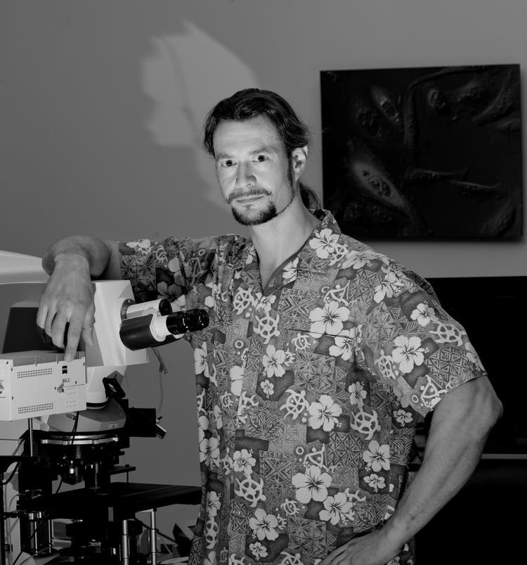
Igor Siwanowicz, Americas winner in Evident’s sixth Annual Image of the Year contest
How did you find the sample you used to create this image?
We have several flowerbeds on campus, and mallows seem to be a part of the wildflower seed mix, so finding the specimen was the easiest part. However, I had to time the collection carefully. When the flowers open, it’s the anthers (male parts) that mature and appear first. It takes the stigma several days to develop its long, white tendrils.
Did you face any challenges when creating this image?
The process wasn’t particularly challenging since I used techniques I’m quite familiar with. It’s a bit like cooking a moderately elaborate dish for the nth time.
Why did you choose this image as your entry for the Image of the Year competition?
I think the image evokes familiar associations—perhaps a sunrise or sunset—while simultaneously depicting something alien or otherworldly. Hopefully, that makes it engaging for a broader audience, and perhaps even slightly confusing. Ideally, though, this is the good kind of confusion—the kind that leads to learning. This is sometimes referred to as “optimal confusion,” and it is thought to be a key epistemic emotion because it drives knowledge acquisition. That kind of confusion creates a certain degree of discomfort—a mental itch that comes with recognizing a gap in one’s knowledge. That itch can only be scratched by finding the information needed to close that gap.
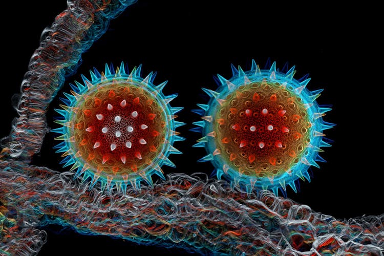
Mallow pollen from the same specimen batch as the winning image for the Americas. Captured by Igor Siwanowicz.
Is there a message inspired by this image?
“The beauty of natural forms can be found on every scale. The right tools help us discover that the beauty of a common flower extends beyond what we can see with the naked eye. It would serve us well not to take nature for granted.”
— Igor Siwanowicz, Americas winner of Evident’s sixth Annual Image of the Year contest
When did you first learn to use a microscope?
My background is in biochemistry and molecular biology. During my PhD, I used NMR and X-ray crystallography to study protein interactions at the atomic level. I felt I was missing the bigger picture. About 19 years ago, I decided to shift my research focus to something more aligned with my interests and joined a neurobiology lab studying memory formation, consolidation, and retrieval using the fruit fly as a model organism. Eventually, after moving to HHMI’s Janelia Research Campus, I carved out my own niche as a specialist in invertebrate anatomy and microscopy.
When did you become inspired to use microscopes to create art?
An early exposure to illustrated textbooks played a huge role. I browsed through them even before I learned how to read. That’s when I first encountered the work of Ernst Haeckel. His Art Forms in Nature, a magnificent collection of highly detailed lithographs showing all sorts of life forms, is a great example of the marriage of scientific approach and artistic talent and remains one of my most influential inspirations.
How long have you been creating art with a microscope?
Around 18 years ago, I collected my first dataset with a confocal microscope (just for fun) shortly after my transition to neurobiology.
What do you find most fascinating about microscopy?
Microscopy images can awaken viewers to the beauty of natural forms and the many facets of design that can be found in nature. They have the power to ignite interest and curiosity, even in non-experts, and motivate some passionate investigations into the natural world. They are a perfect medium for science communication.
On a personal level, microscopy perfectly complements macro photography, something I’ve been doing for the past 20 years, and it gives me an even more intimate perspective on natural forms.
Where do you think this fascination stems from?
My curiosity and interest in the matters of biology came long before any thoughts of an actual career. Both nature and nurture contributed to it in my case. Perhaps there is a genetic factor involved, since both of my parents were biologists.
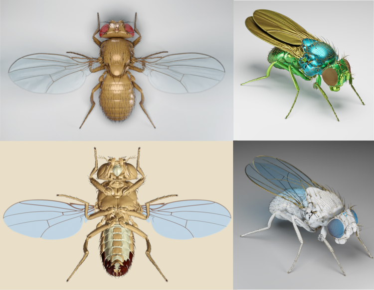
Renders from the anatomical fruit fly model that Igor is currently developing. Captured by Igor Siwanowicz.
What do you do professionally?
I recently added 3D modeling and animation to my toolbox. During the COVID-19 pandemic, I learned a bit of Blender, a free open-source 3D creation suite. It turned out that there is some demand for my new skill, both inside and outside my workplace.
We generate a large number of 3D datasets from imaging techniques such as light microscopy—including confocal and light sheet microscopy—as well as micro-computed tomography (µCT) and FIB-SEM tomography. This data can easily be converted into 3D meshes and imported into Blender for further processing and rendering. Using this approach, I am presently constructing anatomical models of a zebrafish larva, a fruit fly, a bee, a pea aphid, and a mouse.
Does your professional work in imaging intersect with your own artistic work?
There is a considerable overlap between my professional and personal interests. I’d say they’ve pretty much converged, which is the best possible scenario. Besides that, I have developed certain techniques and protocols for imaging various bits of invertebrate anatomy just for fun. They’ve turned out to be very useful in my work.
What kind of experience do you have with Evident microscopes?
I generated my very first “artsy” microscopy images on a FLUOVIEW™ FV1000 microscope, and one of them won first place in Evident’s 2010-2011 BioScapes Imaging Competition. It’s a long story, but I most likely wouldn’t be at Janelia Research Campus right now if that hadn’t happened. My association with the Evident brand and its contests is very positive.
I see a tremendous value in bringing the beauty of nature in all its manifestations to the broader public. I hope the winning images will spark interest and evoke feelings of awe and wonder, which are both particularly vital states of mind in our human experience.
Discover More Beautiful Natural Forms Under the Lens
Want to see more of Igor’s microscope art from the natural world?
Last year, Igor was selected as the global winner of Evident’s fifth Annual Image of the Year contest for his striking cross-section image of a Cosmic Orange Mexican aster flower.
To discover more microscopic images that blend scientific discovery with art, check out our Image of the Year Gallery (don’t miss Igor’s images from 2022 and 2021!)
Plus, be sure to follow Evident on Instagram and LinkedIn to hear about more winner stories and future Image of the Year opportunities.
View Recent Press Coverage
- EVIDENT Announces 6th Annual Image of the Year Award Winners—News Medical
- 17 Award-Winning Microscope Photos Reveal the World’s Hidden Wonders—PetaPixel
- EVIDENT Announces 6th Annual Image of the Year Award Winners—AZoM
- Neuronal Cosmos Wins 6th Annual Image of the Year Award—LABMATE
- Microscopy & Analysis Magazine
- Amazing Winners of This Scientific Microscopic Imaging Contest Capture the Unseen Beauty of Life
- EVIDENT Announces 6th Annual Image of the Year Award Winners—The Scientist
- See 17 Intricate Microscope Photographs That Make the Miniature World Immense. They Won the Evident Image of the Year Contest—Smithsonian Magazine
- International Contest Highlights the World’s Best in Scientific Microscopic Imaging—Wiley Analytical Science
- Germany - Image of the Year Award 2026 - Bildergalerie mit allen im Wettbewerb ausgezeichneten mikroskopischen Aufnahmen—LABO
- Microscopic Art and Celadon Fragments Reveal Hidden Beauty—Artmag
- Korea - Illuminate the Unseen: EVIDENT 제6회 올해의 이미지 어워드—Naver Blog
- Korea - Illuminate the Unseen: EVIDENT 제6회 올해의 이미지 어워드—KakaoTalk
- China - EVIDENT公布第六届年度图像大赛获奖名单!—Weixin