SilVIR-Ray Detector
Fast, Multicolor Super-Resolution. Built on SilVIR Technology.
The SilVIR-RAY™ super-resolution detector extends Evident’s proven SilVIR™ detector technology into image scanning microscopy. Its SilVIR detector array combines super-resolution with low-noise and high-dynamic-range detection, enabling fast, simultaneous multicolor imaging on the FLUOVIEW™ FV5000 confocal and multiphoton laser scanning microscope.
- Product Status: For Research Use Only. Not for use in diagnostic procedures.
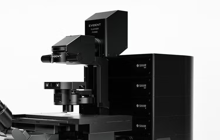
SilVIR-RAY Detector
Multicolor Super-Resolution Built for Speed
SilVIR-RAY™ super-resolution detectors bring SilVIR™ technology into an array designed for image scanning microscopy (ISM). By capturing spatial information that conventional confocal detection leaves behind, SilVIR-RAY detectors enable fast, sensitive super-resolution imaging while retaining the wide dynamic range and low-noise performance the SilVIR detector family is known for.
Available on FLUOVIEW™ FV5000 confocal and FV5000MPE multiphoton laser scanning microscopes, SilVIR-RAY detectors turn super-resolution imaging from an occasional, carefully staged acquisition into something you can use as part of your everyday workflow, even on live, light-sensitive samples.
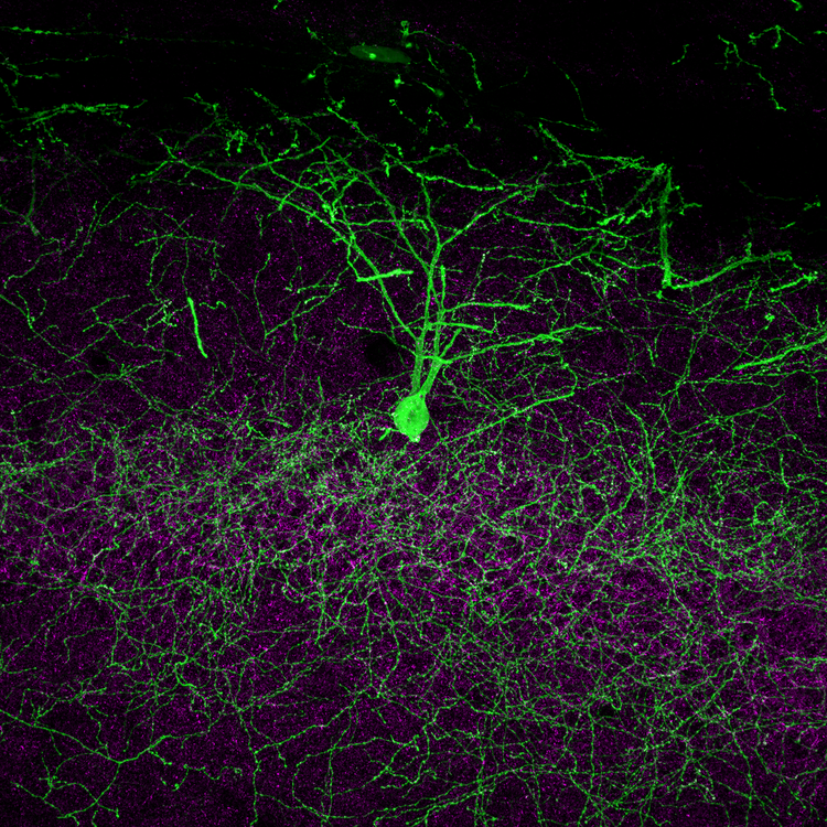
Mouse brain section, hippocampal CA1 region. AAV-labeled neurons with GFP and VGAT immunofluorescence. 8K image acquired with a 25X oil immersion objective (1.00 NA, WD 1.0). Sample courtesy of: Drs. Hirotaka Asai and Shigeo Okabe, Laboratory for Imaging Neural Dynamics, RIKEN Center for Brain Science (CBS).
Explore the technology
Nine SilVIR Detection Elements. One Super-Resolved Image.
SilVIR-RAY reconstructs a super-resolved image in three steps:
1. Collects light and detail at once
A conventional confocal pinhole balances light collection against resolution—open it to collect more light or close it for finer detail. The SilVIR-RAY detector's silicon photomultiplier (SiPM) array does both: multiple sub-pinholes preserve the detail, while together they collect the light of a full Airy unit aperture. This is image scanning microscopy (ISM).
2. Reassembles in real time
Each detector element records a slightly shifted view of the same point source. The SilVIR-RAY detector's patented delay-line pixel reassignment electronically compensates for these shifts in real time before image data are stored—so you get live, high-speed, up-to-4-color super-resolution without flooding your PC with data.
3. Reveals the finest detail
Evident’s TruSight™ SR algorithm maximizes the detail captured by the optical system, providing sharp, high-contrast images through advanced image restoration. TruSight™ deconvolution applies iterative processing to reveal even finer structural detail, enabling resolutions of up to 80 nm in XY and 200 nm in Z. The SilVIR-RAY detector's wide dynamic range ensures those details remain visible across both bright and dim signals in the same acquisition.
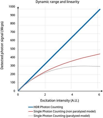
Built on SilVIR Detector Technology
What makes the array architecture particularly effective is the detector technology behind each element. Every element of the array is a SilVIR detector, combining low noise with a wide dynamic range, higher sensitivity, and improved photon-resolving capabilities across the full array.
Quantitative imaging across a wide dynamic range
Imaging data is output as the number of photons, providing the absolute value of the fluorescence intensity for each image. The wider dynamic range provides accurate quantification of fluorescence intensity by photon number even at high intensity levels.
Low-noise detection
Preserve the faint fluorescence signals that demanding super-resolution imaging depends on.
Consistent detection
Semiconductor-based SilVIR technology supports stable detection characteristics across measurements, users, and time points.
Super-Resolution for Dynamic Imaging
SilVIR-RAY detectors perform spatial reassignment as the signal is acquired, avoiding the need to generate and store multiple separate detector images for every super-resolved frame. This reduces the data-processing burden and enables super-resolution imaging with the FV5000's fast scanning modes.
Behind this capability is delay-line pixel reassignment, which performs spatial correction at the electrical-signal stage rather than reconstructing the image afterward from nine separately stored images.
High-resolution 3D volume rendering of apical dendrites from a Thy1-YFP H line mouse brain section, with dendritic spine heads clearly visualized. Objective: UPLAPO100XOHR (100X oil, NA 1.50). Sample courtesy of: Dr. Tetsushi Hoshida and Prof. Atsushi Miyawaki, RIKEN Center for Brain Science (CBS).
Simultaneous acquisition preserves both temporal fidelity and the true spatial relationship between structures. COS-7 cells labeled with βtublin-mStayGold (microtubules) and EB1-mScarlet, showing protein localization at the cell periphery. Courtesy of Yasushi Okada, University of Tokyo.
See Multiple Targets at the Same Moment
Up to four SilVIR-RAY detection units can operate simultaneously on a single FV5000 platform, enabling simultaneous multicolor super-resolution acquisition. Different fluorescence channels are captured at the same time, avoiding the temporal offset of sequential acquisition.
Delay-line pixel reassignment makes this practical by consolidating each detector array's signal into a single image as it is acquired, reducing the data volume that would otherwise be generated by multiple array detectors.
Super-Resolution That Preserves Signal Relationships
Higher resolution is only useful if you can trust it. The SilVIR-RAY detector's super-resolved images are reconstructed using Evident’s TruSight™ SR algorithm, a linear image-processing framework extended to support ISM.
Both the underlying processing and the SilVIR-RAY detector’s response are linear, so the reconstructed image remains proportional to the fluorescence signal of your sample.
The result is imaging that’s not just visually sharper, but quantitatively meaningful.
TruSight SR also lets you continuously adjust processing strength to match the signal-to-noise of your image, so low-signal, live-cell data doesn't have to be sacrificed to get a sharper picture.
Learn more about TruSight SR technology
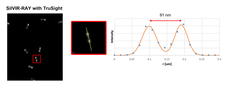
SilVIR-RAY + TruSight images of a nanoruler showing 91 nm resolving power.
From Fine Structures to Dynamic Processes
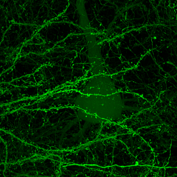
Dendritic arbor imaged at high speed without loss of structural detail. Soma and branching processes captured across an 80 μm depth range at 0.1 μm Z-steps, using a 100X objective (UPLXAPO100XO) in 2K resonant scan mode with 10x accumulation—demonstrating that resonant scanning preserves fine structural fidelity even across large, information-dense fields of view.
Explore the Technology Behind SilVIR-RAY
Go deeper into the array detector architecture, delay-line pixel reassignment, SilVIR detection characteristics, super-resolution processing, and performance validation.
FLUOVIEW FV5000
SilVIR-RAY on the FLUOVIEW FV5000
SilVIR-RAY detectors integrate with the FLUOVIEW FV5000 platform, combining super-resolution detection with the system's confocal imaging, scanning, automation, and workflow capabilities. Up to four SilVIR-RAY detection units can be installed on a single FV5000 system, alongside its standard SilVIR detector channels.
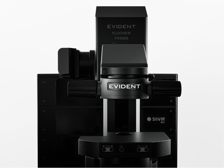
The FLUOVIEW FV5000 confocal and multiphoton laser scanning microscope.