Uncovering Microscopic Landscapes—6th Annual Image of the Year Materials Science Winner
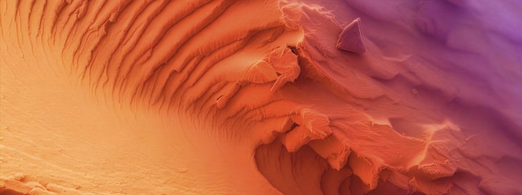
Evident Image of the Year Award 2025: Materials Science Winner.
Lignin fiber captured by Muhammad Tahir Khan, Ireland.

For Muhammad Tahir Khan, the materials science winner of Evident’s sixth annual Image of the Year contest, the laboratory is a gateway for not just understanding materials but seeing them differently.
While doing his PhD research at the University of Limerick, Ireland, Muhammad uncovered the hidden world of lignin, a key structural component of plant cell walls. Usually associated with rigidity and resistance, this image reveals another identity of lignin—one defined by elegance, depth, and subtle beauty.
Muhammad was selected as the materials science winner for his image “The Ember Dunes of Lignin.” What appears as dunes are actually the result of chemistry: molecular interactions shaped by processing, structure, and the intrinsic complexity of lignin itself. The image was captured using a scanning electron microscope.
In recognition of his winning submission, he will receive an Evident SZ61 stereo microscope. Muhammad shared insights on how he captured an image that evokes both the beauty of an unexplored landscape and the hidden universe under the microscope lens.
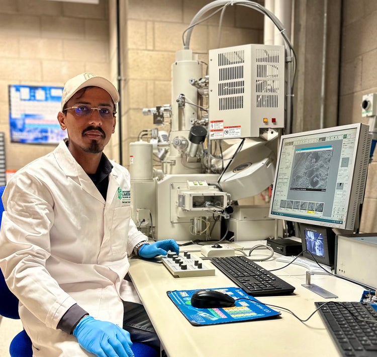
Muhammad Tahir Khan, of Ireland, was named the materials science winner in Evident’s sixth annual Image of the Year competition.
Muhammad, congratulations on being named the Image of the Year materials science winner! What did you find personally exciting about this image?
This image reminds me of the similarities and versatility of the universe, from the micro to the macro level. Sometimes, a tiny fragment that we can hold at the tip of our finger can resemble something that is worth billions of dollars and takes years to create or discover.
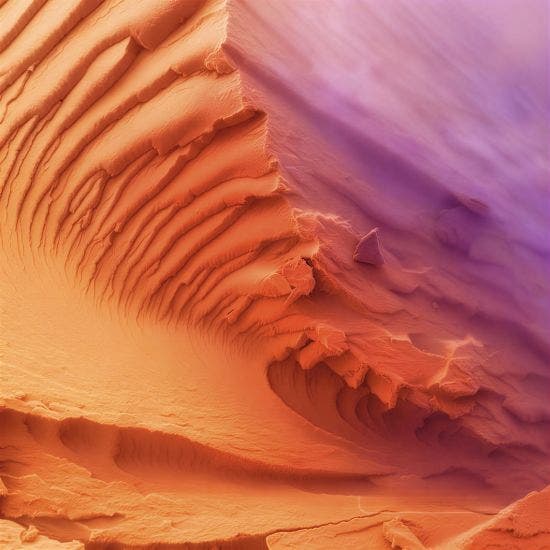
Evident Image of the Year Award 2025: Materials Science Winner.
Lignin fiber captured by Muhammad Tahir Khan, Ireland.
How did you create the image?
The image was captured using a scanning electron microscope at the Bernal Institute, University of Limerick, Ireland. The samples used to create the image are part of my PhD research and were developed at the Bernal Institute, University of Limerick, Ireland, under the CARBOWAVE project funded by Horizon Europe.
Did you face any challenges when creating this image?
Scientific work always involves a certain level of difficulty. For these specific samples, the extremely humid Irish weather posed a significant challenge, as some components are highly hydrophilic, which is an undesirable characteristic for the intended application.
Why did you choose this image as your entry for the competition?
Initially, I was examining the material interface without expecting anything extraordinary. As I explored the sample further, the surface and its ridge-like structures appeared remarkably fascinating, resembling an unexplored landscape. At that moment, I knew this image was something special.
Is there a message inspired by this image?
“Strength is not always visible. It is built layer by layer beneath the surface.”
— Muhammad Tahir Khan, materials science winner of Evident’s sixth annual Image of the Year contest
When did you first learn to use a microscope?
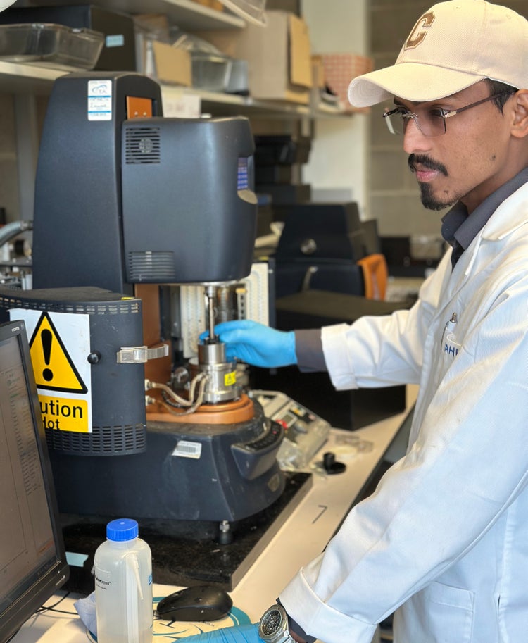
I first started using microscopes during my master’s studies and later had the opportunity to work extensively with various types of microscopes. I have a strong scientific background, having completed both my undergraduate and postgraduate studies in polymer engineering, along with over five years of professional experience in academic research and industry.
When did you become inspired to use microscopes to create art?
I have always been an explorer. Initially, I was fascinated by the stars and deeply curious about what lies beyond the sky. During high school, I came across striking micrographs of natural structures, which inspired me to explore the universe at a much smaller scale. That moment marked the beginning of my journey into microscopy, which gradually became one of my favorite pursuits.
A defining turning point came last year when I saw the winning images from an annual scientific imaging competition at the University of Limerick. The winning photograph was displayed on the institution’s wall of fame, which left a strong impression on me. It motivated me to further develop my microscopy skills, with the goal of one day seeing my own work displayed there.
How long have you been creating art with a microscope?
I’ve been involved in this field for the past 10 years, but it was only last year that I began creating art through microscopy.
What do you find most fascinating about microscopy?
Microscopy is fascinating because it reveals hidden worlds beyond human perception. It turns tiny structures into meaningful insights, showing how structure explains function. At the same time, it blends science with art, transforming the invisible into something both understandable and visually striking.
Where do you think this fascination stems from?
It stems from a mix of curiosity and discovery—the human drive to see beyond limits and understand how things truly work. Microscopy satisfies that by revealing hidden structures, turning the unknown into something visible, meaningful, and often unexpectedly beautiful.
What do you do professionally?
I am a PhD student in the School of Engineering at the University of Limerick, Ireland.
Does your professional work in imaging intersect with your own artistic work?
Yes, very much. My work in imaging often naturally overlaps with artistic expression. The microstructures I study for scientific purposes frequently resemble landscapes or abstract art, so the same images that provide technical insights also inspire creative interpretation. It’s a space where analysis and aesthetics meet. I am not just studying materials but also appreciating and uncovering their hidden beauty.
What kind of experience do you have with Evident microscopes?
I have experience operating several Evident microscopes, including stereo optical microscopes and digital microscope systems, which I found to be smooth, reliable, and user-friendly.
See More Images That Illuminate the Unseen
Want to see more microscopic images where science meets art? Explore our Image of the Year Gallery. You can also follow Evident on Instagram and LinkedIn to stay up to date on future Image of the Year opportunities.
View Recent Press Coverage
- EVIDENT Announces 6th Annual Image of the Year Award Winners—News Medical
- 17 Award-Winning Microscope Photos Reveal the World’s Hidden Wonders—PetaPixel
- EVIDENT Announces 6th Annual Image of the Year Award Winners—AZoM
- Neuronal Cosmos Wins 6th Annual Image of the Year Award—LABMATE
- EVIDENT Announces 6th Annual Image of the Year Award Winners—The Scientist
- See 17 Intricate Microscope Photographs That Make the Miniature World Immense. They Won the Evident Image of the Year Contest—Smithsonian Magazine
- International Contest Highlights the World’s Best in Scientific Microscopic Imaging—Wiley Analytical Science
- Germany - Image of the Year Award 2026 - Bildergalerie mit allen im Wettbewerb ausgezeichneten mikroskopischen Aufnahmen—LABO
- Korea - Illuminate the Unseen: EVIDENT 제6회 올해의 이미지 어워드—Naver Blog
- Korea - Illuminate the Unseen: EVIDENT 제6회 올해의 이미지 어워드—KakaoTalk
- China - EVIDENT公布第六届年度图像大赛获奖名单!—Weixin
Featured Products
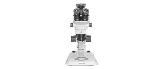
SZ61/SZ51
Research Stereo Microscope System
The compact SZ61 and SZ51 stereo microscopes deliver flat, natural color images, smooth macro-to-micro-observation, and generous depth of field for routine life science, production, assembly, and inspection tasks.
- SZ61: magnification range of 6.7x–45x, zoom ratio of 6.7:1
- SZ51: magnification range of 8x–40x, zoom ratio of 5:1
- Greenough optical system for excellent image flatness, large depth of field, and excellent stereoscopic view
- High color fidelity for observing and documenting specimens in natural colors
- Reflected and transmitted LED illumination options for wide-ranging observation
- Antistatic construction and an available trinocular SZ61 model for digital imaging
- Wide choice of auxiliary objectives to modify magnification from 2x to 270x and a working distance up to 350 mm