Microscopic Art from the Heart—6th Annual Image of the Year APAC Winner
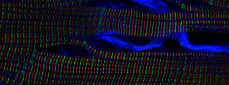
Evident Image of the Year Award 2025: APAC Winner.
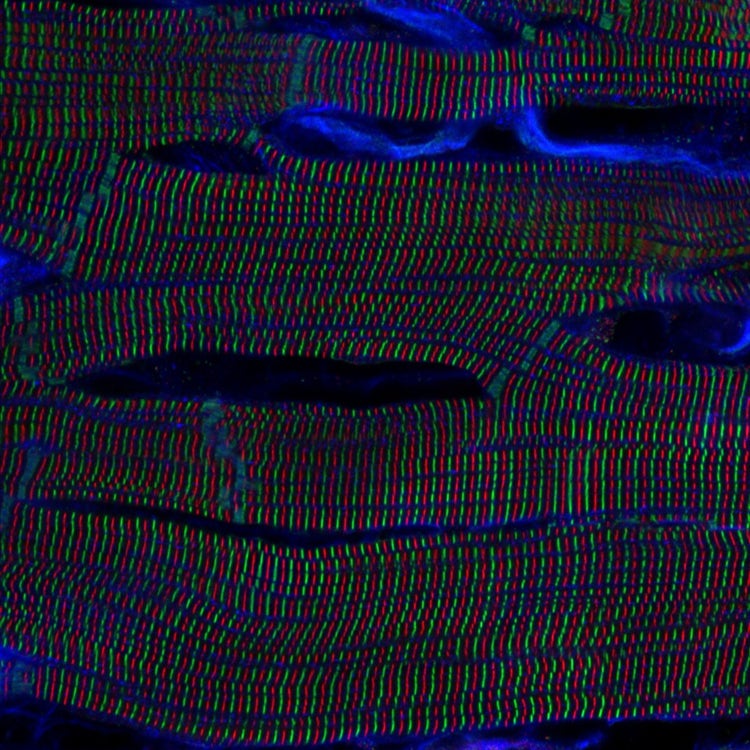
Sarcomere structures within cardiomyocytes in a rat heart. Captured by Kentaro Mochizuki, Japan.

A precisely ordered network of green and red lines reveals the microscopic architecture behind every heartbeat. Captured by Dr. Kentaro Mochizuki of Japan, this image of sarcomere structures in rat cardiac muscle cells was named the APAC winner of Evident’s sixth annual Image of the Year competition.
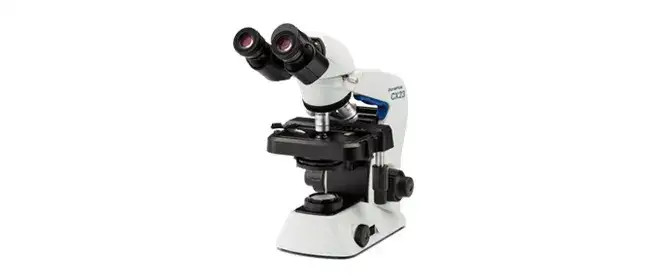
Using a confocal laser microscope and a 63X oil immersion objective, Kentaro brought the heart’s linear, rhythmic microstructure into vivid focus. In recognition of his winning submission, he will receive an Evident CX23 upright microscope.
Illuminating the Unseen Structures Hidden Within the Heart
As a researcher in the Department of Pathology and Cell Regulation (PCR) at Kyoto Prefectural University of Medicine, Kentaro spends his days studying living organisms and disease. Through confocal microscopy, he reveals structures that are invisible to the naked eye—including the orderly patterns within cardiac muscle that make each heartbeat possible.
Evident Image of the Year Award 2025: APAC Winner.
Sarcomere structures within cardiomyocytes in a rat heart. Captured by Kentaro Mochizuki, Japan.
Kentaro, congratulations on being named the Image of the Year APAC winner! What do you find personally exciting about this image?
The orderly alternation of Z-lines (green) and M-lines (red) that form the sarcomere structure of cardiac muscle cells evokes a sense of both mysterious beauty and primal fear, drawing one in unconsciously.
The heart pumps blood throughout the body by continuously repeating the cycle of contraction and relaxation, more commonly known as the heartbeat. This beating is generated when every single cell that makes up the heart muscle contracts and relaxes in unison. In sync with this movement, the spacing between the Z-lines and M-lines also narrows and widens. As you observe this image, place your hand on your chest to feel your heartbeat, and imagine how, with each beat, this periodic structure pulsates like a spring.
How did you create the image?
Perfused rat hearts were chemically fixed with paraformaldehyde (PFA), sectioned into tissue blocks measuring several millimeters square, and stained with fluorescent dyes. Z-lines (green) and M-lines (red) were labeled via immunofluorescence staining for α-actinin and titin, respectively, while the cell membrane region (blue) was labeled using wheat germ agglutinin (WGA).
For imaging, the stained tissue sections were placed on a glass slide with a mounting medium, and a confocal laser microscope was used. This image was captured using a 63X oil immersion objective (NA 1.4), showing an in-plane region (88 µm × 88 µm) of the subepicardial myocardium as viewed from the epicardial side.
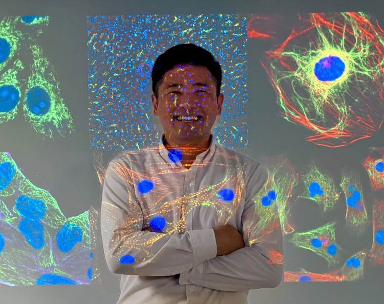
Dr. Kentaro Mochizuki, of Japan, was named the APAC winner in Evident’s sixth annual Image of the Year competition.
How did you find the sample you used to create this image?
I actually used this sample in my own cardiac research. Although this image will not be published in a paper, it is a piece of data that holds special significance for me because it provided crucial guidance during the early stages of my research. I am truly delighted to have this opportunity to share it in this way.
Did you face any challenges when creating this image?
We encountered several technical challenges during the staining and imaging processes. To preserve the microstructure of the myocardium as naturally as possible, we stained and imaged the tissue as whole sections without performing sectioning. During the staining process, we focused on membrane permeabilization to ensure the antibody could reach deep within the tissue, as well as on optimizing antibody concentration and reaction time.
While using the optical sectioning effect of the confocal microscope, we paid meticulous attention to the positioning of the sample and focus adjustment to ensure that the outermost layer of myocardial cells on the curved heart surface remained within the imaging plane.
Why did you choose this image as your entry for the Image of the Year competition?
I wanted to share the beauty and mystery of the sarcomere structure in the heart muscle with as many people as possible. When you hear the term “microstructure of living organisms,” you might imagine something curvilinear and irregular, but I hope you’ll see that linear, orderly, and periodic structures actually exist within the body. I would also be delighted if this opportunity sparks even a little interest in the world of research.
Is there a message inspired by this image?
While I was deeply moved by the intricate periodicity of cardiac sarcomeres, I also felt compelled to explore whether any heterogeneity might be hidden within this orderly structure. In recent years, various forms of heterogeneity have been discovered in biological phenomena and structures, and research into their significance and importance is progressing—the heart is no exception.
For example, I hope to elucidate the spatiotemporal balance with which this periodic structure expands and contracts across the entire tissue during cardiac contraction, thereby contributing to our understanding of complex pathologies such as heart failure.
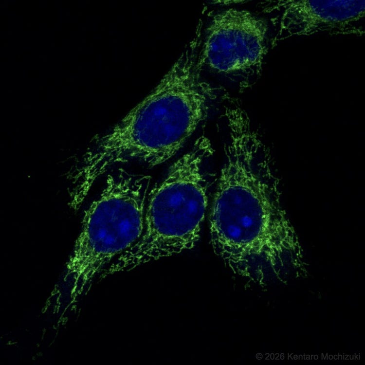
Fluorescent immunostaining image of HeLa* cells captured using an Evident FV1000 confocal microscope.
*To learn more about the origin of HeLa cells, visit henriettalacksfoundation.org.
When did you first learn to use a microscope?
I first used a professional microscope shortly after being assigned to my first research lab as a senior in college. Through that experience, I learned not only to acquire microscope images as experimental data, but also to appreciate the beauty of the images and the biological structures themselves. Having grown up surrounded by nature from a young age, I had already developed the habit of carefully observing plants and animals with the naked eye or a magnifying glass.
When did you become inspired to use microscopes to create art?
I think it was the first time I saw the fine structures inside cells (probably mitochondria and lipid droplets) through a fluorescence microscope. Rather than creating art, I felt as though a fully realized work of art was already unfolding before my eyes as I peered through the microscope, so I captured and savored that beauty within the bounds of my research.
How long have you been creating art with a microscope?
It’s been about three years. As I’ve had more opportunities to perform immunostaining and microscopic observation of cells and tissues for my research, I’ve also become more meticulous about screening—that is, determining which antibodies to use under what conditions to clearly visualize the microstructures I’m studying (essentially obtaining positive controls).
During the screening process, I sought out the optimal angles and perspectives to bring out the full detail of each cell and tissue. Whenever I captured a shot I was satisfied with, I would admire it or show it to a colleague sitting next to me to get their feedback. Looking back now, it feels as though I was photographing cells and tissues while having them strike various poses.
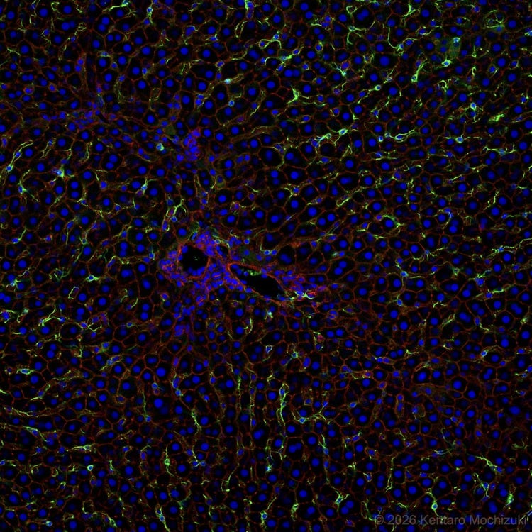
Fluorescent immunostaining image of a rat liver captured using an Evident FV1000 confocal microscope.
What do you find most fascinating about microscopy?
“What fascinates me most is the ability to visualize things that are invisible to the naked eye. Even after understanding the principles and science behind them, the various microscopy techniques—which do more than simply magnify small objects but also make invisible objects visible—still feel almost magical to me.”
— Dr. Kentaro Mochizuki, APAC winner of Evident’s sixth annual Image of the Year contest
Where do you think this fascination stems from?
I have always loved nature and living things, and I have a strong curiosity about how they are structured and function, rather than their overall appearance. This passion is reflected in my current research, where I uncover the hidden world of living organisms and diseases. The world of cells and tissues viewed through the microscope is a fascinating blend of dynamic chaos and stillness as well as structural roughness and delicacy—a world that never ceases to captivate me and continues to inspire my enthusiasm for both research and art.
What do you do professionally?
I am a researcher in the Department of Pathology and Cell Regulation (PCR) at Kyoto Prefectural University of Medicine (KPUM). My background is in engineering, and within the field of biomedical engineering, I work daily to solve biological and medical challenges by using optical technologies, including microscopy.
Does your professional work in imaging intersect with your own artistic work?
In my daily research activities, I am engaging with the art that cells and tissues naturally create. In addition to sharing the resulting microscope images with my colleagues, I have recently begun sharing microscope images—which, though not published in papers, possess academic value or artistic merit—on X and Instagram as part of the “PositiveControlSharing” series, with the aim of sharing information with other researchers and giving back to society.
What kind of experience do you have with Evident microscopes?
I have been using Evident microscopes since I was first assigned to the lab about 16 years ago. In addition to the successive models of the inverted IX™ series and the upright BX™ series, I have recently been using the MVX10 macro zoom microscope system frequently for research purposes. I also continue to use the FV1000 confocal laser scanning microscope––which has been in our lab for over 10 years—while taking great care to maintain it.
View Recent Press Coverage
Explore additional coverage of Evident’s sixth annual Image of the Year competition to see how researchers and imaging enthusiasts around the world are using microscopy to uncover striking structures in cells, tissues, and the natural world. Plus, follow Evident on Instagram and LinkedIn for future winner stories, competition updates, and opportunities to share your own remarkable microscope images.
- EVIDENT Announces 6th Annual Image of the Year Award Winners—News Medical
- 17 Award-Winning Microscope Photos Reveal the World’s Hidden Wonders—PetaPixel
- EVIDENT Announces 6th Annual Image of the Year Award Winners—AZoM
- Neuronal Cosmos Wins 6th Annual Image of the Year Award—LABMATE
- EVIDENT Announces 6th Annual Image of the Year Award Winners—The Scientist
- See 17 Intricate Microscope Photographs That Make the Miniature World Immense. They Won the Evident Image of the Year Contest—Smithsonian Magazine
- International Contest Highlights the World’s Best in Scientific Microscopic Imaging—Wiley Analytical Science
- Germany - Image of the Year Award 2026 - Bildergalerie mit allen im Wettbewerb ausgezeichneten mikroskopischen Aufnahmen—LABO
- Korea - Illuminate the Unseen: EVIDENT 제6회 올해의 이미지 어워드—Naver Blog
- Korea - Illuminate the Unseen: EVIDENT 제6회 올해의 이미지 어워드—KakaoTalk
- China - EVIDENT公布第六届年度图像大赛获奖名单!—Weixin
Featured Products
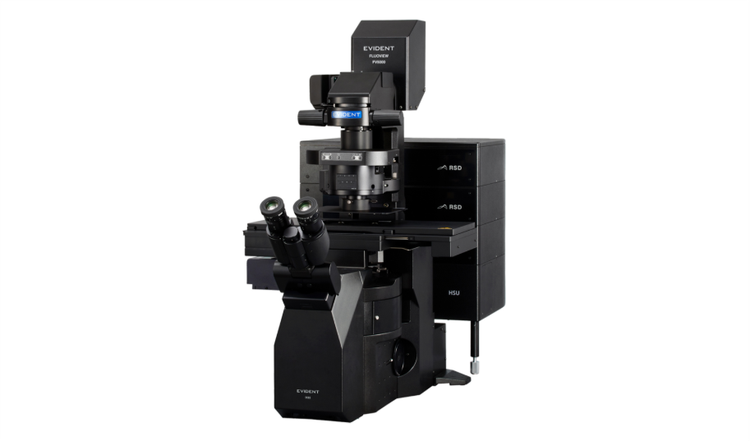
FV5000
Confocal Laser Scanning Microscope
- Extraordinary clarity, speed, and reliability driven by groundbreaking innovations
- SilVIR™ detectors deliver photon-level quantitation, exceptional sensitivity, and ultra-high signal-to-noise
- Unmatched dynamic range captures the full signal spectrum and prevents saturation
- High-speed 2K resonant scanning and high-density 8K galvo scanning in one platform
- FLUOVIEW Smart™ software simplifies operation with intuitive controls and AI-powered automation
- TruResolution™ auto correction collar optimizes focus for over 20 objectives
- Modular design supports up to 10 laser lines and future multiphoton upgrades
- Laser Power Monitor (LPM) ensures stable illumination and reproducible results over time