Improving Reproducibility in Confocal Microscopy at Shared Imaging Facilities
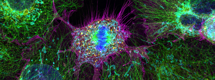
Multichannel confocal image of cultured cells. Instrument-layer automation helps maintain consistent imaging conditions across users and sessions.

Reproducibility in confocal microscopy depends on more than sample preparation and user technique. Instrument performance, illumination stability, detection sensitivity, acquisition settings, and metadata capture can all vary between sessions.
These variables are especially difficult to control at shared imaging facilities, where multiple researchers with different levels of experience use the same instrument. A more consistent approach begins by identifying which conditions can change and moving their measurement and control closer to the instrument.
For a more elaborate explanation and a practical facility assessment, download the white paper, Lock Down Confocal Reproducibility Before Users Touch the Scope.
Why Reproducibility Is Difficult at Shared Imaging Facilities
Reproducibility is difficult at shared imaging facilities because different users, changing instrument conditions, and incomplete metadata can all affect the final image. Facility staff must support researchers with varying levels of experience yet cannot observe every setup decision or verify every acquisition condition in real time.
A 2022 UK study, cited in the downloadable white paper, found wide variance in core facility management, with the same two-person staff responsible for anywhere from 20 to 280 users. This makes manual supervision difficult, particularly when variability may not become apparent until images are compared across sessions, users, or sample batches.
The documentation challenge is just as significant. A 2020 review of 240 research articles, also referenced in the paper, found that only 17.7% of fluorescence microscopy papers met minimum standards for reporting acquisition parameters and equipment metadata. Important details can therefore be missing from the published record even when the resulting images appear suitable for analysis.
Reproducibility begins before the first image is captured. It depends on whether the system can measure and control the conditions that shape the image, rather than relying entirely on user memory, training, and manual documentation.
Why Laser Power and Software Settings Aren’t Enough
Software percentages describe a selected setting, not necessarily the amount of laser output delivered to the sample. A session log that records a laser setting of 10% documents a dial position, but it does not confirm how much illumination reached the sample during acquisition.
One study, quoted in the downloadable white paper, described laser output variation of up to 50-fold across confocal systems from the same vendor. Within a single three-hour session, solid-state lasers commonly fluctuate by 25% to 50%, according to the same report.
The same software setting can therefore represent different illumination conditions between instruments or across sessions. For quantitative imaging, a recorded percentage isn’t enough to explain an intensity difference. The relevant question is not only what setting the user selected, but what output the instrument actually delivered at the time of acquisition.
This distinction is the starting point for instrument-level reproducibility: separating the software command from the instrument’s measured performance.
Why Reproducibility Needs to Move to the Instrument Layer
Reproducibility should move to the instrument layer because shared facilities cannot rely on user training and manual oversight alone to control every condition that affects an image. Routine microscope maintenance and checks to confirm that the system is operating within its expected parameters also place a significant time burden on facility staff.
Instrument-layer quality control can support several parts of the imaging process:
- Illumination: A laser power monitoring system can measure laser output against an installation baseline and support correction when delivered power changes.
- Instrument performance: A microscope performance monitoring system can assess laser power, detection sensitivity, and imaging performance on a scheduled basis.
- Detection: Photon-resolving detection can provide a more reliable basis for comparing intensity across sessions when the optical configuration remains consistent.
- Workflow setup: Automated tools can help standardize sample positioning, focus, laser power adjustment, and startup settings for rotating users.
Variation introduced at any one of these layers can propagate into the resulting data, even when the other conditions are well-controlled. Together, these capabilities can support acquisition conditions that remain comparable across sessions and users on the same system.
How Does the FLUOVIEW™ FV5000 Support Reproducible Imaging?
The Evident FLUOVIEW™ FV5000 confocal laser scanning microscope supports reproducible imaging through photon-count quantitation, laser drift correction, scheduled performance monitoring, and automated workflow tools in compatible configurations.
Its SilVIR™ detectors support photon-level quantitation, while the Laser Power Monitor helps maintain consistent laser output across sessions. The Microscope Performance Monitor evaluates system sensitivity and imaging consistency, helping facility staff identify changes in instrument performance within a few minutes. FLUOVIEW Smart™ software provides automated tools that can help simplify routine setup for users with different levels of experience.
Learn more about FLUOVIEW Smart software and its approach to intuitive, AI-enhanced confocal workflows.
For a closer look at how SilVIR supports quantitative confocal imaging, read the white paper: Next-Generation SilVIR Detector System for the FLUOVIEW FV5000 Laser Confocal Microscope.
Build a Stronger Reproducibility Baseline
Confocal reproducibility depends on controlling the conditions that shape an image, not only reviewing the image after acquisition. Laser output, detector behavior, optical performance, setup decisions, and metadata all contribute to the record associated with an experiment.
Instrument-layer automation can make calibration, performance monitoring, acquisition setup, and reporting part of the baseline workflow. For shared imaging facilities, that approach can help researchers work with greater confidence across sessions while giving facility managers clearer evidence when an image or experiment requires investigation.
Download the white paper, Lock Down Confocal Reproducibility Before Users Touch the Scope, to see how these layers work together and benchmark your facility with the eight-point readiness checklist.
Featured Products
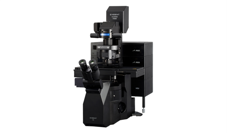
FV5000
Confocal Laser Scanning Microscope
- Extraordinary clarity, speed, and reliability driven by groundbreaking innovations
- SilVIR™ detectors deliver photon-level quantitation, exceptional sensitivity, and ultra-high signal-to-noise
- Unmatched dynamic range captures the full signal spectrum and prevents saturation
- High-speed 2K resonant scanning and high-density 8K galvo scanning in one platform
- FLUOVIEW Smart™ software simplifies operation with intuitive controls and AI-powered automation
- TruResolution™ auto correction collar optimizes focus for over 20 objectives
- Modular design supports up to 10 laser lines and future multiphoton upgrades
- Laser Power Monitor (LPM) ensures stable illumination and reproducible results over time
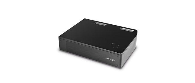
SilVIR Detector
FLUOVIEW Laser Scanning Microscope Solutions
- Combines a silicon photomultiplier and patented * fast signal processing for lower noise, higher sensitivity, and improved photon resolving capabilities
- High detection efficiency provides superior signal-to-noise to bring weak fluorescence to life
- Capture vivid fluorescence images with no offset adjustments
- Precisely quantify image intensity for more reliable data
*Patent number US11237047