Why Microscope Field of View Matters in Pathology: A Practical Comparison

How does the field of view change your pathology workflow? We compare FN22 and FN26.5 oculars across screening speed, HPF counting, and viewing experience.
Many pathologists work with FN22 oculars because that’s the industry standard. But what if you could see even more?
Increasing the field number to 26.5 gives you 45% more viewable area—a percentage that translates into real workflow differences.
In this guide, we compare the differences between a 22 mm and 26.5 mm field of view, highlighting three key benefits of a larger field of view in pathology workflows.
Comparing a 22 mm vs. 26.5 mm Field of View
In 1975, the optical engineers at Evident introduced a 26.5 mm field of view, a milestone for upright microscopes that’s still industry-leading today.
Compared to FN22, FN26.5 reveals 45% more surface area.
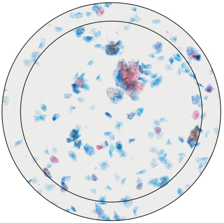
Thin layer of cells. Field of view at 20X magnification for FN22 (inner circle) and FN26.5 (outer circle).
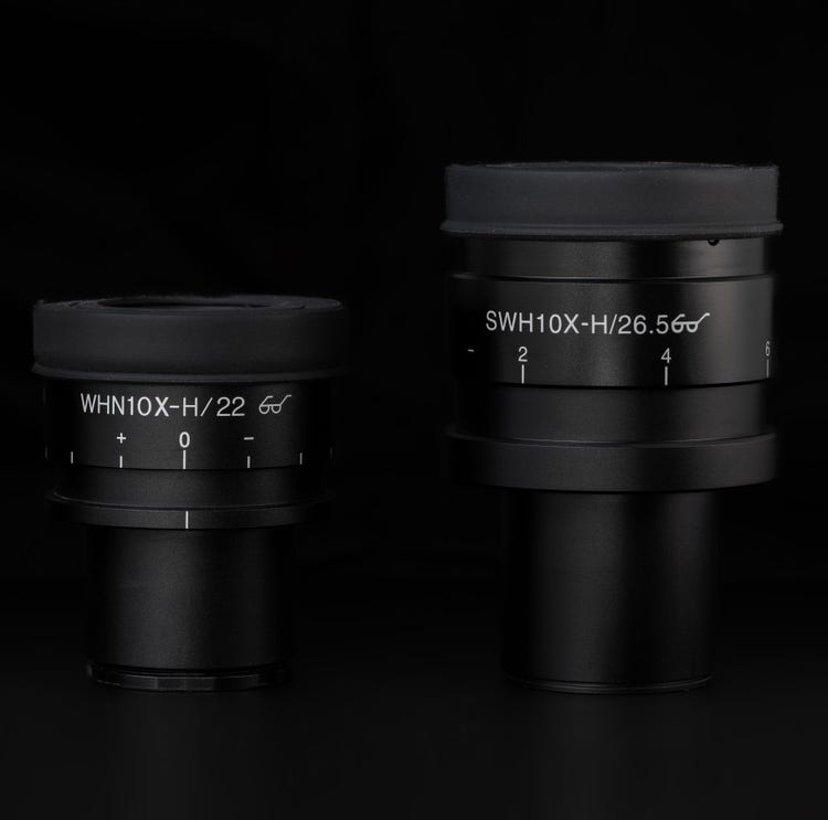
Physical difference between an FN22 ocular (left) and an FN26.5 ocular (right).
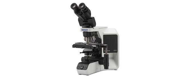
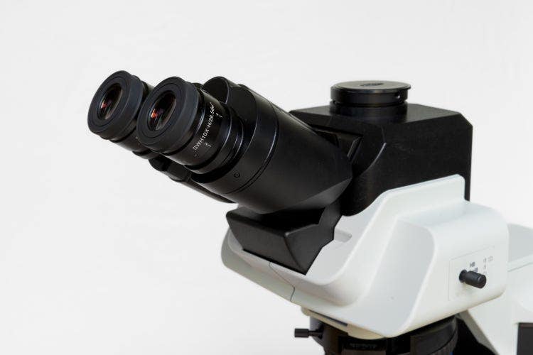
The 26.5 mm field of view is available on Evident BX43 and BX53 upright microscopes, as well as legacy BX™ microscopes that can be upgraded with compatible observation tubes, objectives, and oculars.
Here are three key advantages of switching to a 26.5 mm field of view in pathology:
1. Faster Screening at Low Magnification
Increasing the field of view from the standard 22 mm to 26.5 mm area lets you view 45% more of the slide. This leads to faster initial screening under the microscope.

Here’s an example: At 2X magnification, you need two passes instead of three to see the full slide. At 4X magnification, it’s four passes instead of five. Many pathologists find it to be more efficient, especially at low magnification.
“Using the FN26.5 ultra-wide field observation tube has made me feel that it can effectively enhance work efficiency. The work efficiency of scanning cytology slides at 10x magnification has been greatly improved, whether for cell smears or thin-layer liquid-based slides.”
Associate Chief Physician
Department of Pathology, China
“The super-wide-angle observation head with FN26.5 has been a game-changer in my workflow. Its large field of view makes scanning and reviewing cases at low power incredibly efficient, allowing me, as a histopathologist, to quickly identify areas of interest with minimal eye strain. Combined with high-quality objectives—2X, 4X, 10X, and 20X—the super-wide-angle head supports rapid, confident evaluations without the frequent need to switch to higher magnifications. It’s an ideal setup for anyone looking to streamline their diagnostic process.”
Dr. Gordon Harloe
MB ChB M MED PATH (ANAT) FRCPath FRCPA FAICD
Consultant Anatomical Pathologist
2. A Smarter Way to Count High-Power Fields
A great example showing the advantage of the 26.5 mm field of view at high magnification is when counting high-power fields (HPF). Often, pathologists need to count events on their samples at 40X magnification by evaluating 10 different fields of view and comparing the results with a reference table.
In many cases, the reference is based on 10 HPFs using FN22 oculars, which cover a total sample area of 2.38 mm2. You can explore more details in our application note, HPF Counting Remains a Core Tool in Clinical Microscopy.
In short, FN26.5 can improve HPF counting workflows in two ways:
1. Faster counting with fewer fields to evaluate: Since the field of view is 45% larger than FN22, counting can be completed after only 7 HPFs with a total area of 2.41 mm2—very close to 2.38 mm2 reference area for FN22. This offers a time advantage since less slide movement is required.
2. More flexibility for inhomogeneous samples: For highly inhomogeneous samples, you can still count 10 HPFs and apply a correction factor of 0.69 to make the results comparable to the 2.38 mm2 reference area used with FN22.
When the reference is based on the sample, such as 2 mm2 or 5 mm2 area instead of the 2.38 mm2 area of FN22, use Table 1 to determine the number of HPFs needed.
| Visible area per field number (mm2) | HPF for 2 mm2 | HPF for 5 mm2 | |||||||
| FN20 | FN22 | FN26.5 | FN20 | FN22 | FN26.5 | FN20 | FN22 | FN26.5 | |
| 1.25X | 201.06 | 243.28 | 352,99 | - | - | - | - | - | - |
| 2X | 78.54 | 95.03 | 137,89 | - | - | - | - | - | - |
| 4X | 19.63 | 23.76 | 34,47 | - | - | - | - | - | - |
| 10X | 3.142 | 3.801 | 5,515 | - | - | - | - | - | - |
| 20X | 0.785 | 0.950 | 1,379 | 2.5 | 2.1 | 1.5 | 6.4 | 5.3 | 3.6 |
| 40X | 0.196 | 0.238 | 0,345 | 10.2 | 8.4 | 5.8 | 25.5 | 21.0 | 14.5 |
| 60X | 0.087 | 0.106 | 0,153 | 22.9 | 18.9 | 13.1 | 57.3 | 47.4 | 32.6 |
| 100X | 0.031 | 0.038 | 0,055 | 63.7 | 52.6 | 36.3 | 159.2 | 131.5 | 90.7 |
Table 1. Left: visible area of view in mm2 for different magnifications at FN20, FN22, and FN26.5. Middle: number of HPF to cover 2 mm2. Right: number of HPF to cover 5 mm2.
3. A More Comfortable Viewing Experience
Research has shown that visual strain symptoms are common among full-time microscope users. In fact, one ergonomics study reported symptoms in 80% of operators. Therefore, a more relaxed viewing experience from FN26.5 oculars is a meaningful contribution to improving comfort during daily microscope use.
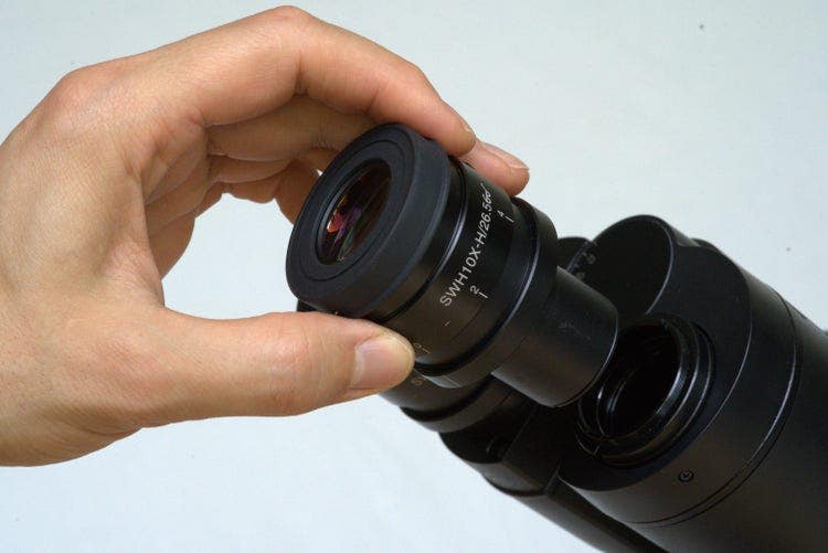
FN26.5 ocular
When working with FN22 oculars, eyes tend to lock onto the center of the field. But with a 26.5 mm field of view, there’s so much more to see. The eyes will start wandering around, exploring the sample. That’s what makes the 26.5 mm view more relaxing. It’s closer to a natural viewing experience. We encourage pathologists and lab professionals to test this wider view for themselves, and many users have reported a similar experience.
"The FN26.5 ocular adds a significant upgrade to my daily BX microscope setup. Its immersive view is easier on the eyes, helps me work faster, and gives me confidence that I’m not missing details—especially at the edges of the slide."
Professor
Department of Dermatology, Venereology, and Allergology
Charité–Universitätsmedizin Berlin, Germany
“I personally really like this ultra-widefield observation tube; I feel that the images under the microscope are much closer to the human eye, creating a lifelike experience. Since my gaze does not need to be focused frequently, it also feels very comfortable for my eyes when reviewing slides.”
Attending Physician
Pathology and Cytology Department, China
Ready to experience the benefits of this wider field of view? If you’d like to know if your current BX system can be upgraded to FN26.5, reach out to us today to book a demo!
Disclaimer: Certain names in the testimonials have been withheld for legal and confidentiality reasons.
Featured Products
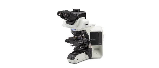
BX53
- High-luminosity True Color LED illumination
- Modular concept enables individual components to be motorized
- Easily acquire multi-color fluorescence images