Image of the Year 2024 di EVIDENT

We are thrilled to announce the winners of our 5th annual Image of the Year contest, recognizing the world’s best in scientific microscopic imaging. This year’s contest included our first-ever video category.
This year’s winners were selected from entries from 29 countries. We thank everyone who entered, and we look forward to your participation again next year.


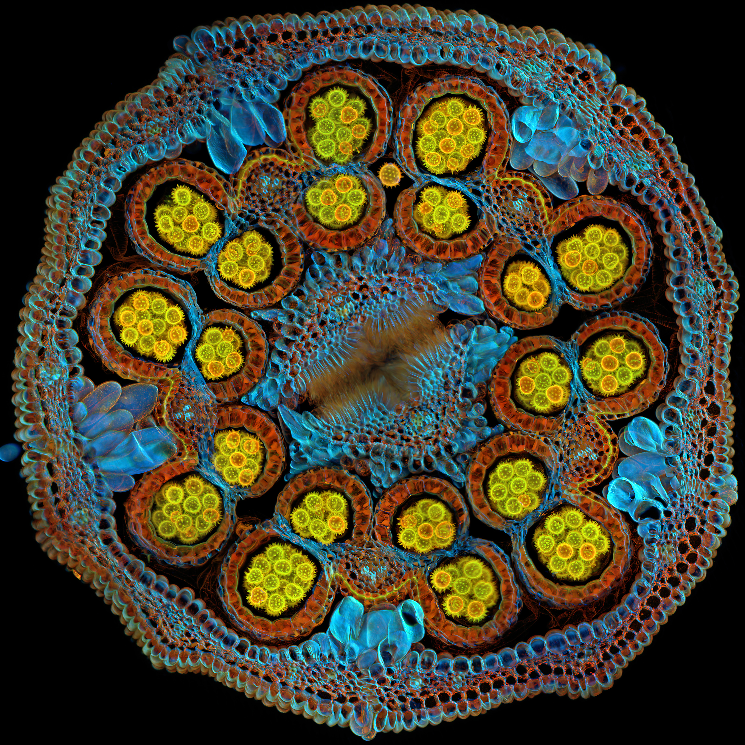
Vincitore globale
Bellezza eccezionale
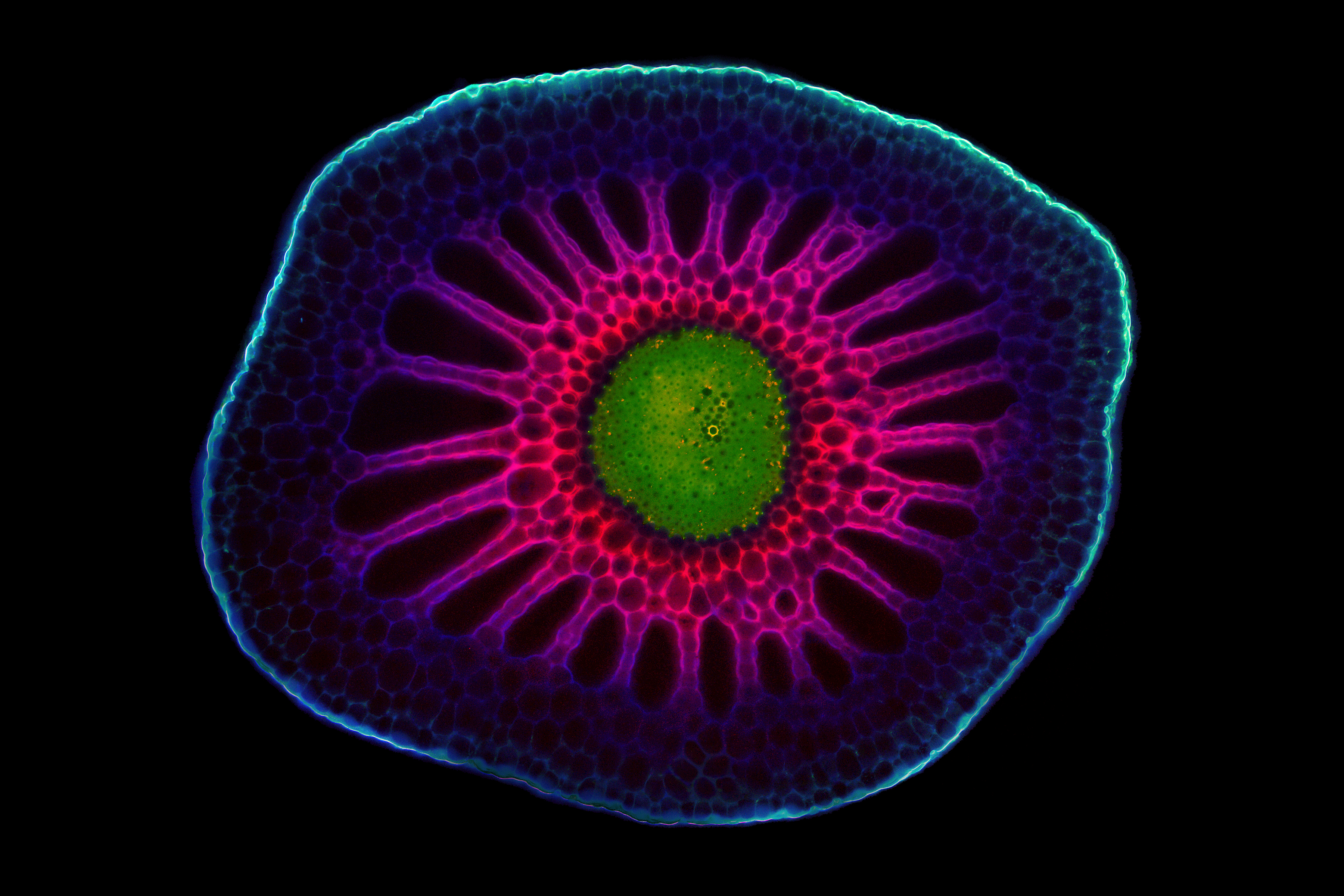
Sezione trasversale di cosmea sulfurea (Cosmos sulphureus) raffigurante la maturazione dei granuli pollinici all'interno delle antere. Immagine acquisita da Igor Siwanowicz (Stati Uniti).
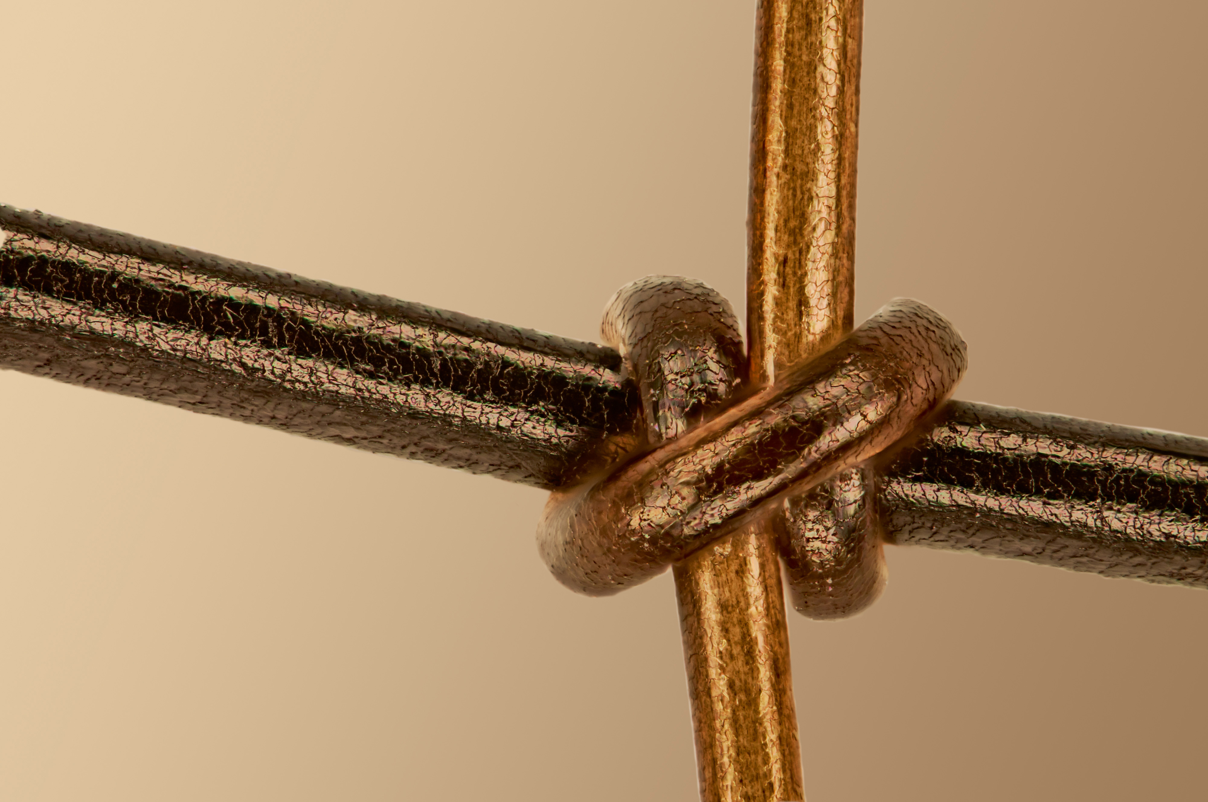
Vincitore per la categoria scienze dei materiali
Connettere tutte le forme viventi
Capello (verticale) annodato a un crine di cavallo (orizzontale). Immagine acquisita da Gerd Günther (Germania).

Vincitore per la categoria video
Un universo vasto e sconfinato
Processo di cristallizzazione di un amminoacido. Acquisito da Zhigang Zheng (Cina).
Americhe

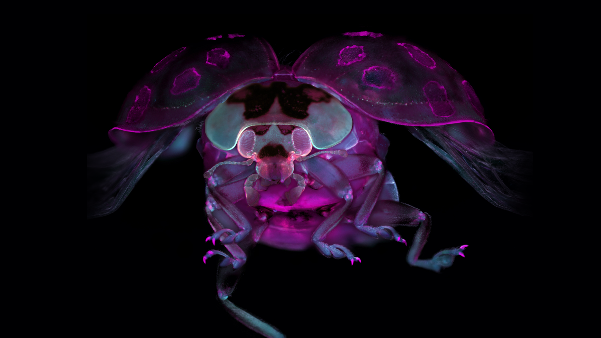
La scoperta di nuove prospettive
Coccinella con tessuto chiarificato. Immagine acquisita da Marko Pende (Stati Uniti).
EMEA

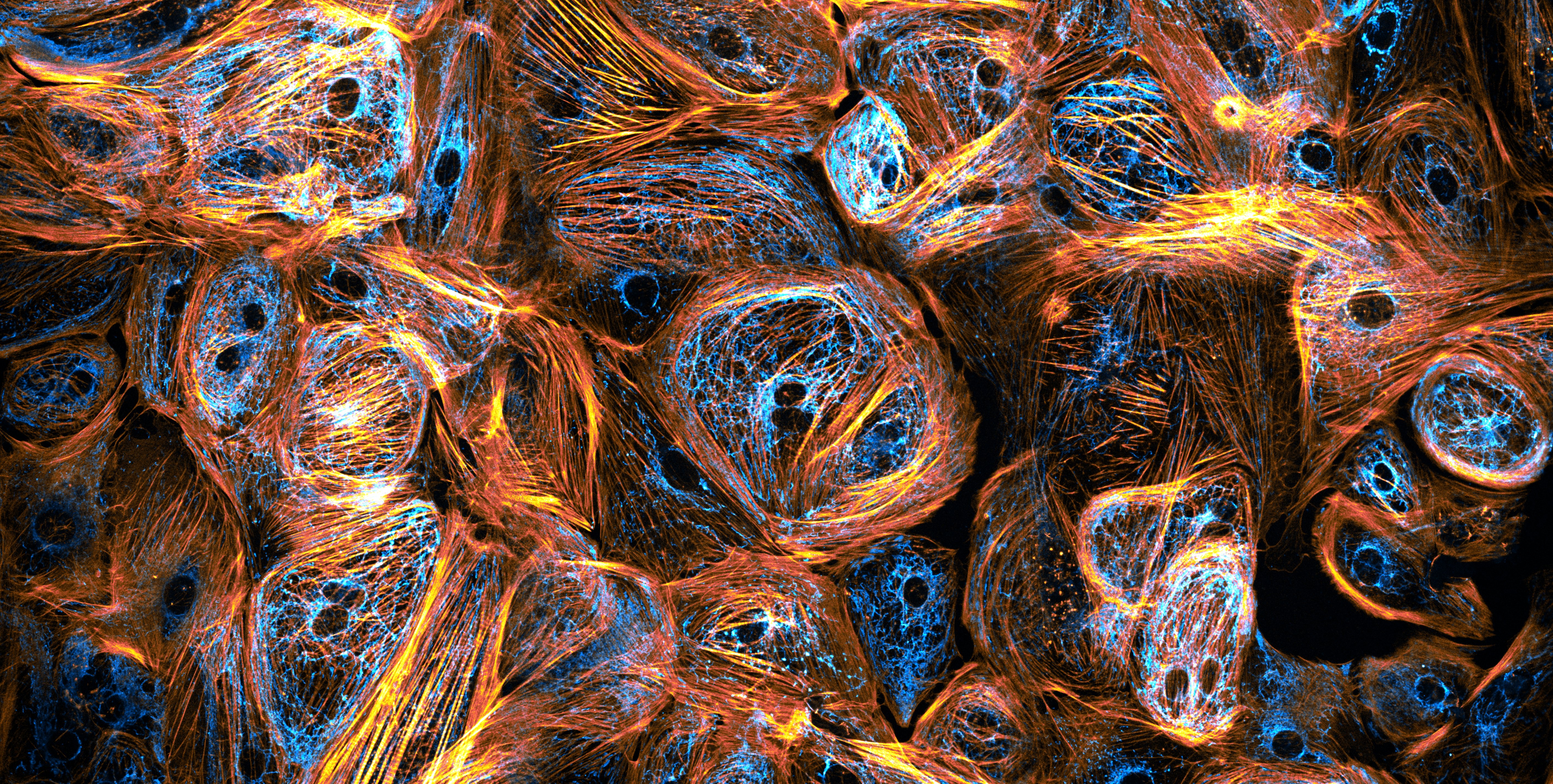
Natura come artista supremo
Cellule muscolari derivate da cellule staminali pluripotenti indotte. Immagine acquisita da Till Stephan (Germania).
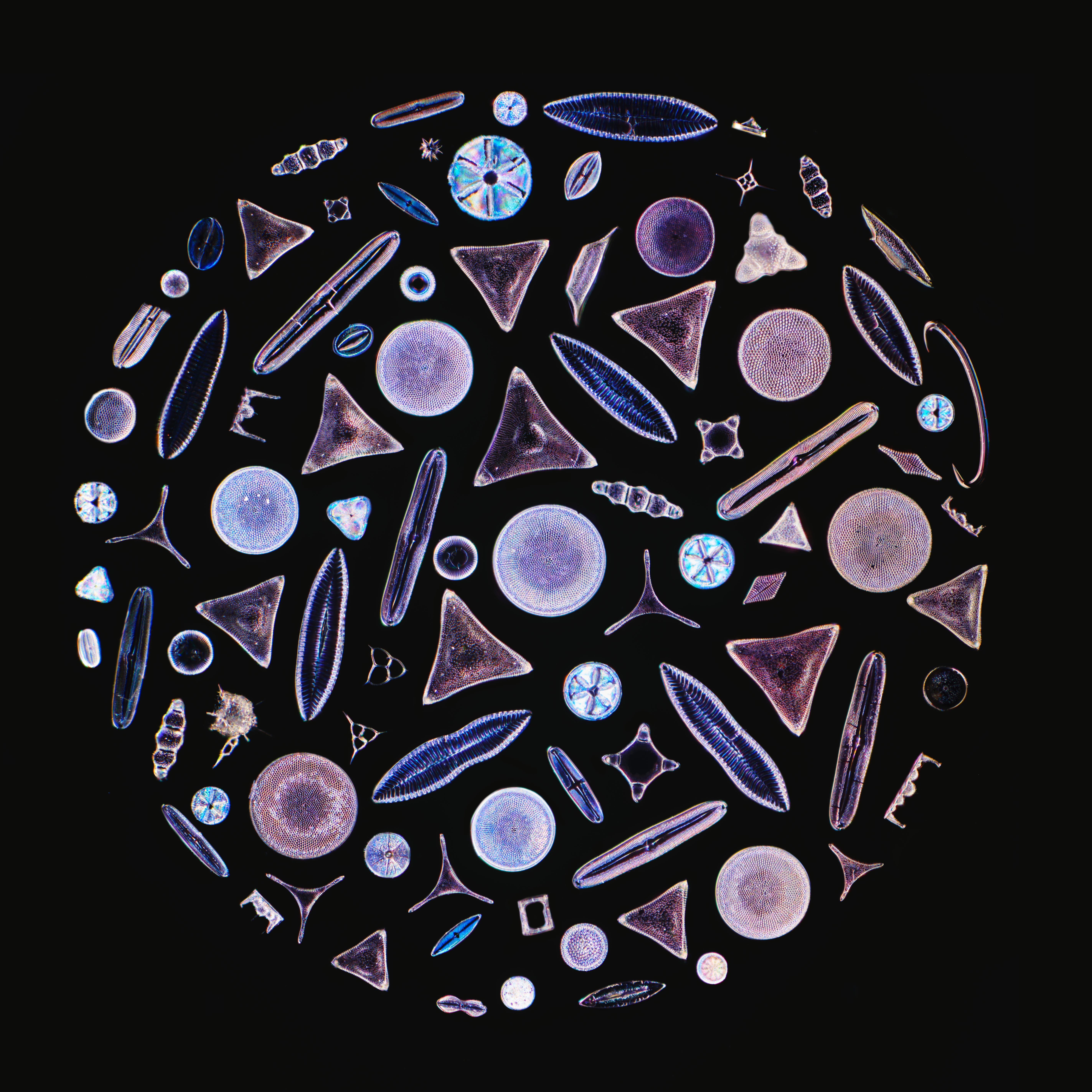
Asia-Pacifico

Incontro con le gemme della natura
Composizione di diatomee.Immagine acquisita da Daniel Han (Australia).
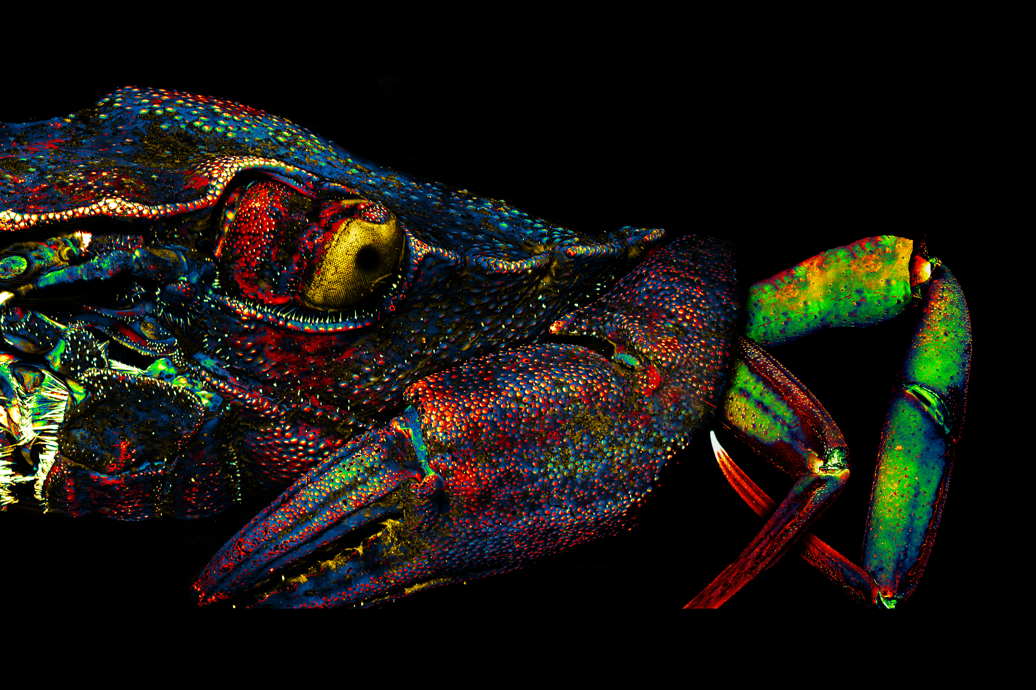
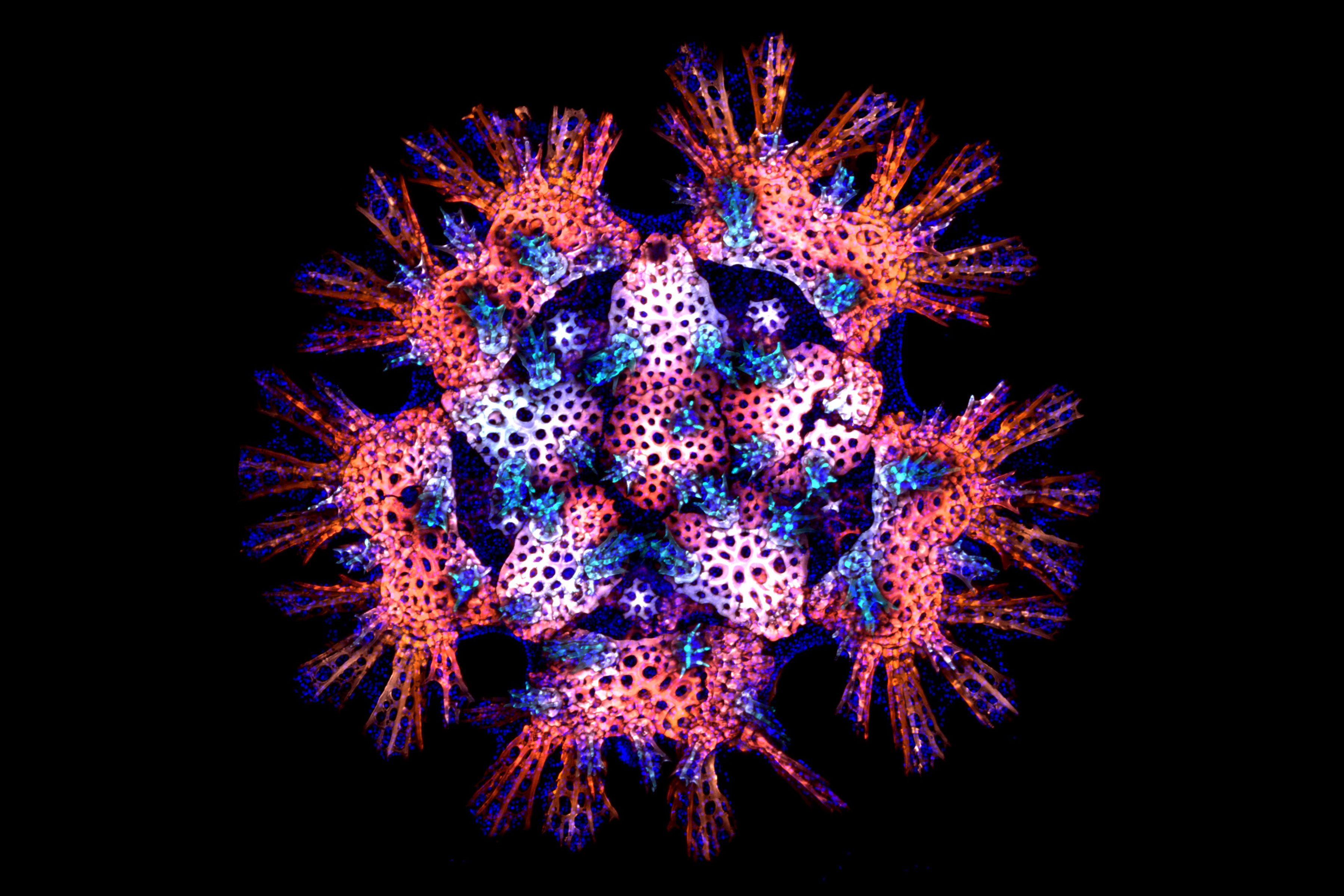
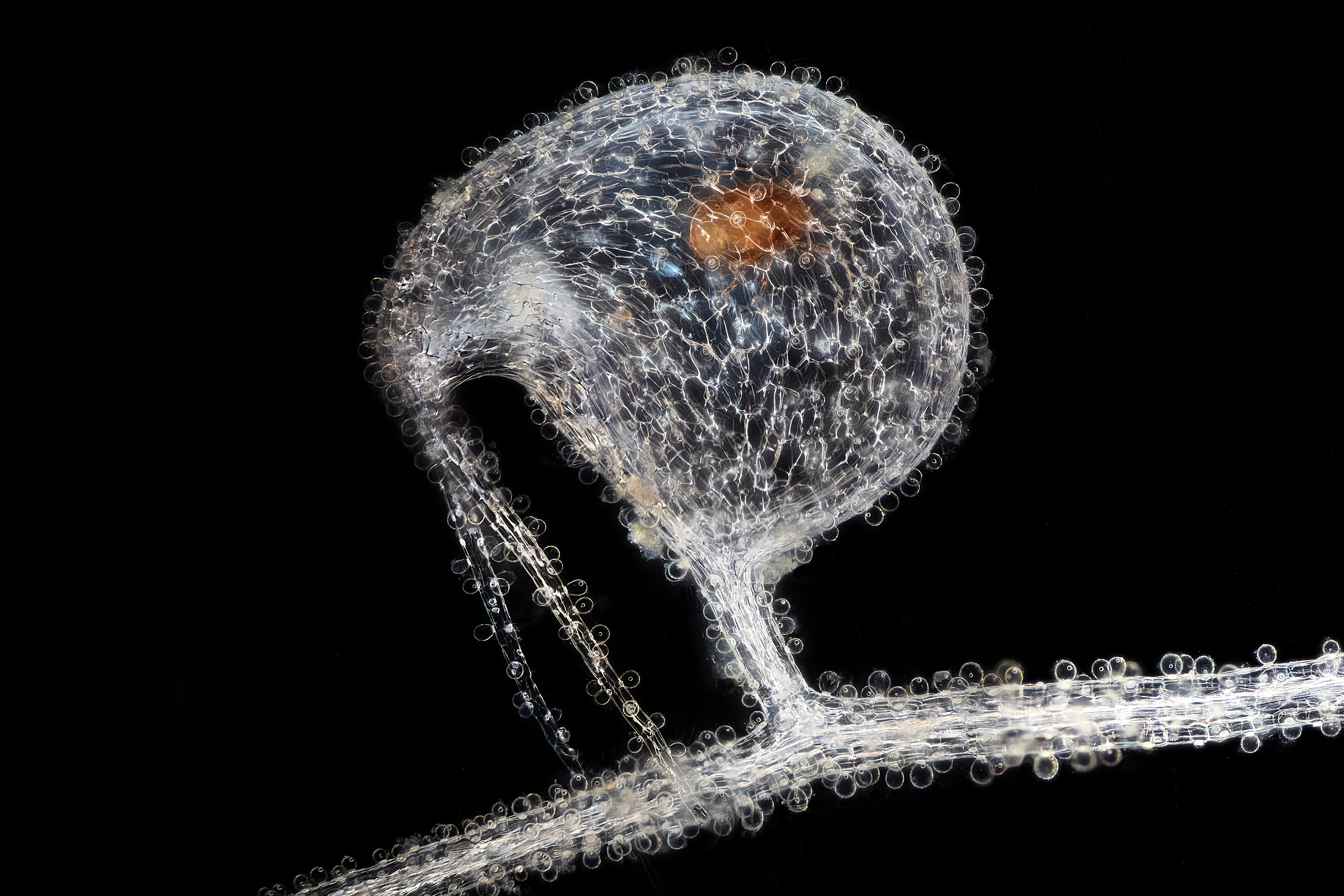
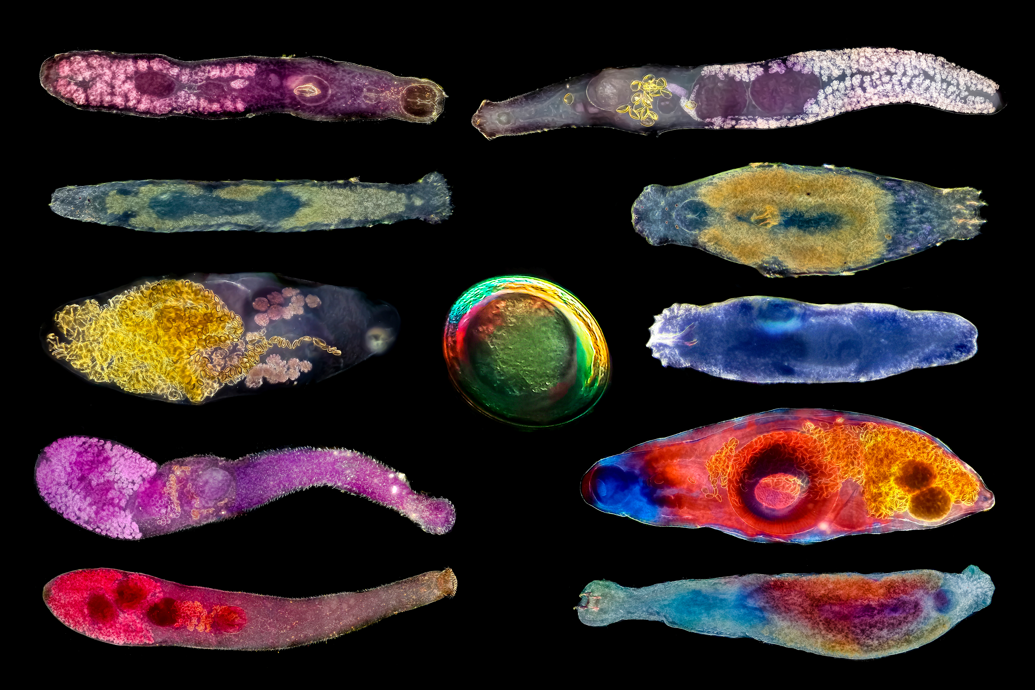
Menzioni speciali

Granchio verde, marcato con DAPI e rappresentato con immagini con autofluorescenza a luce riflessa.

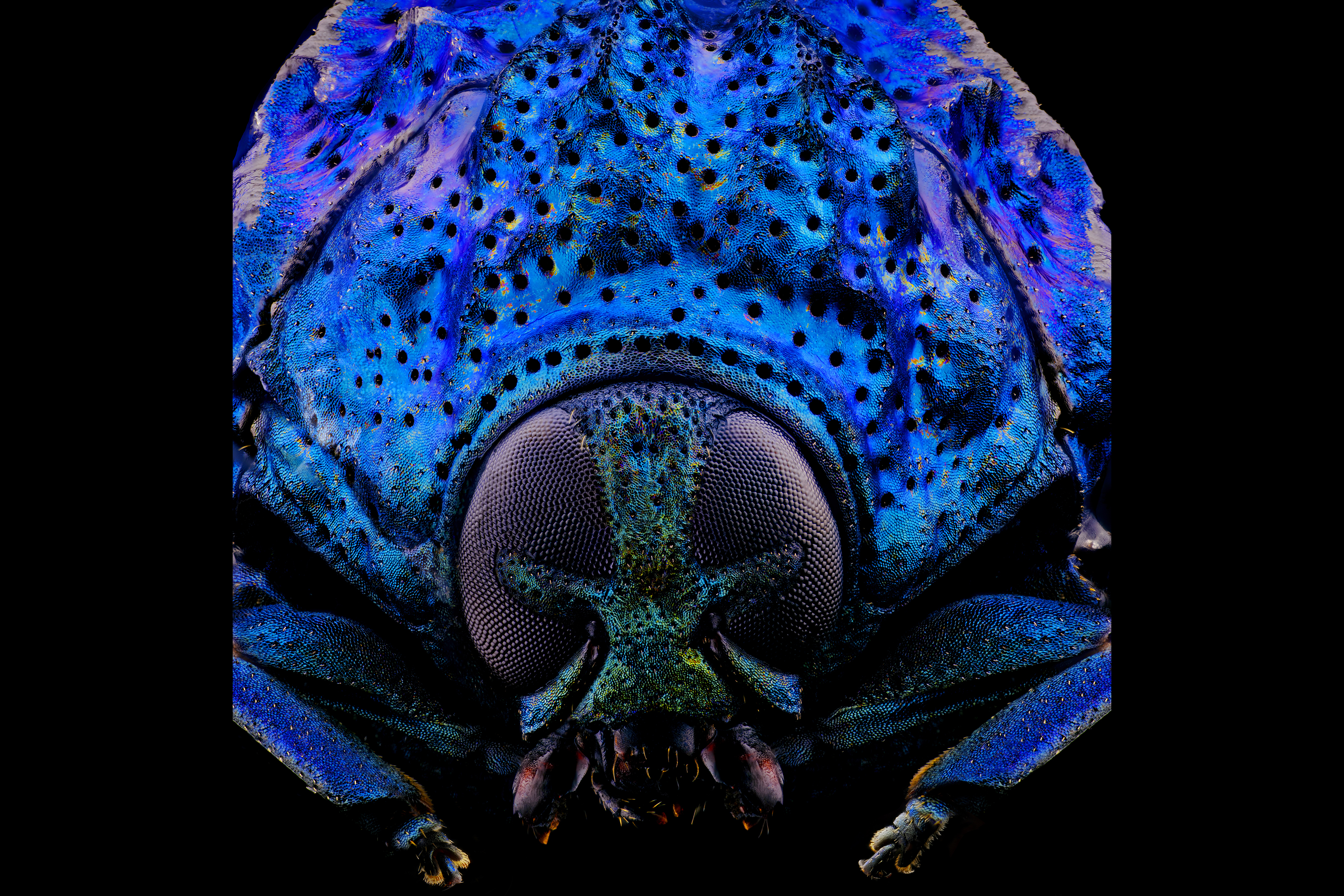
I Crisìdidi sono insetti affascinanti che sfoggiano una meravigliosa gamma di colori iridescenti.

Scheletro di giovane stella di mare. Giovane stella di mare (Patiria miniata) trattata con i coloranti di calceina e DAPI.

Immagine di farfalla costituita da più di 200 motivi con scaglie provenienti da diverse ali di farfalle.

Trappola di urticularia carnivora subacquea (Utricularia humboldtii) con intrappolato un acaro acquatico (Hydrachnidia)

Collage di diversi parassiti di pesci osservati in campo scuro in Patagonia (Argentina).

Un crisomelide (Poropleura) somigliante a un tirannosauro preistorico.

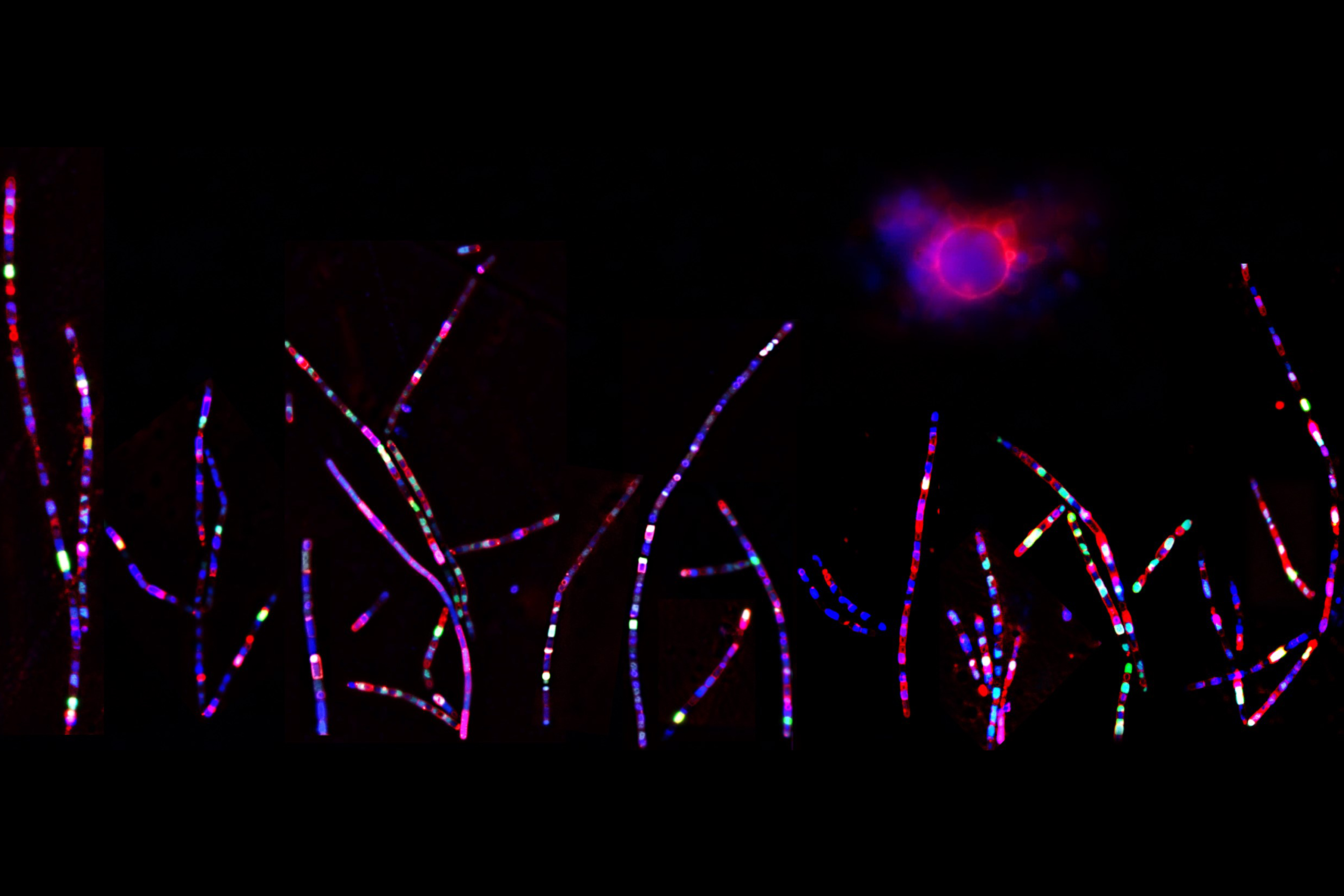
Sandra Story (Stati Uniti)
Immagini fluorescenti di cellule filamentose normali a forma di bastoncello del batterio del suolo Bacillus subtilis e di una cellula sferica che ha perso la parete cellulare.

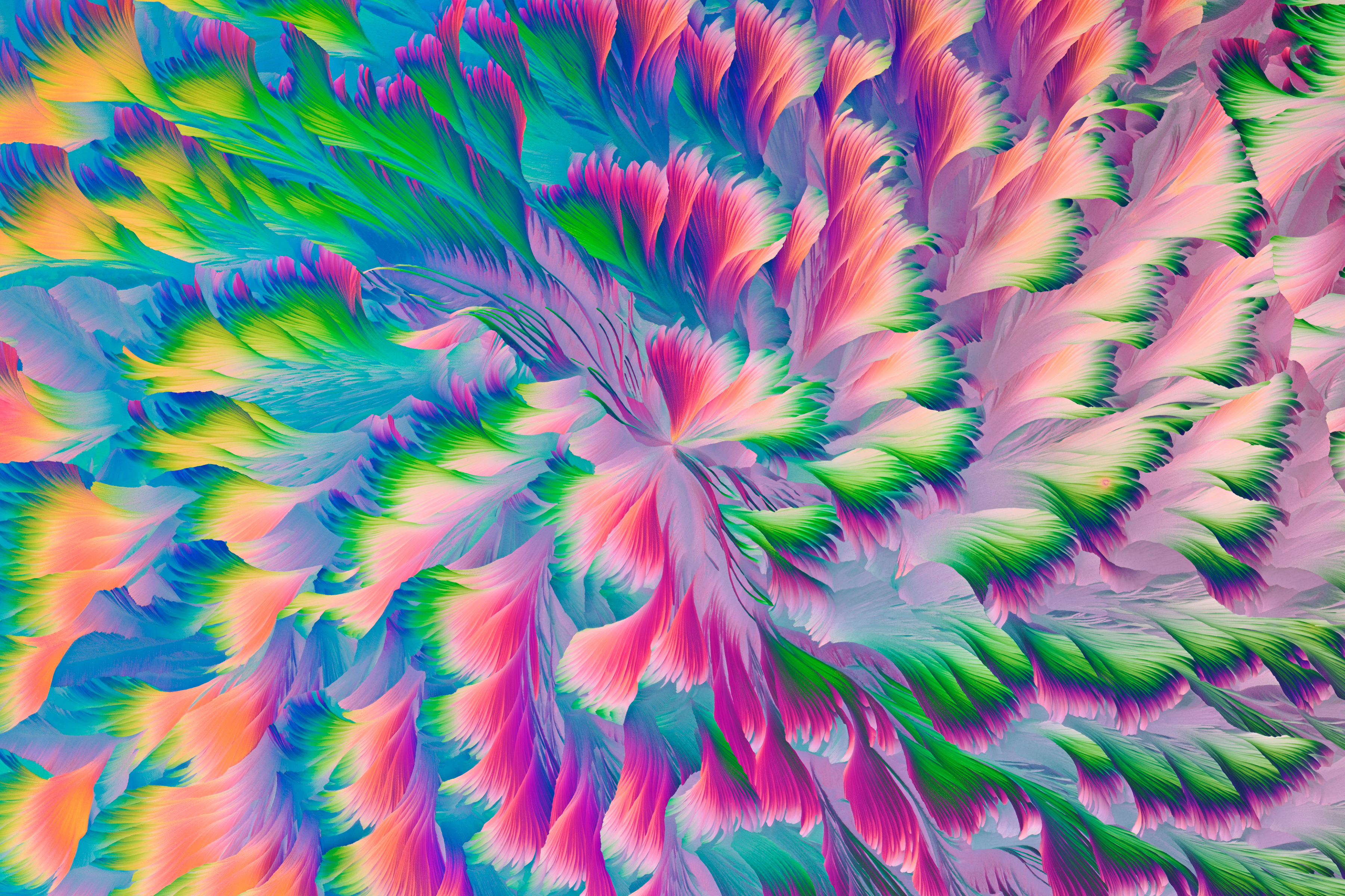
Ji Yuan (Cina)
Motivo cristallino, simile a quello di una grande giacca imbottita in cotone utilizzata in occasione della Festa della Primavera cinese.

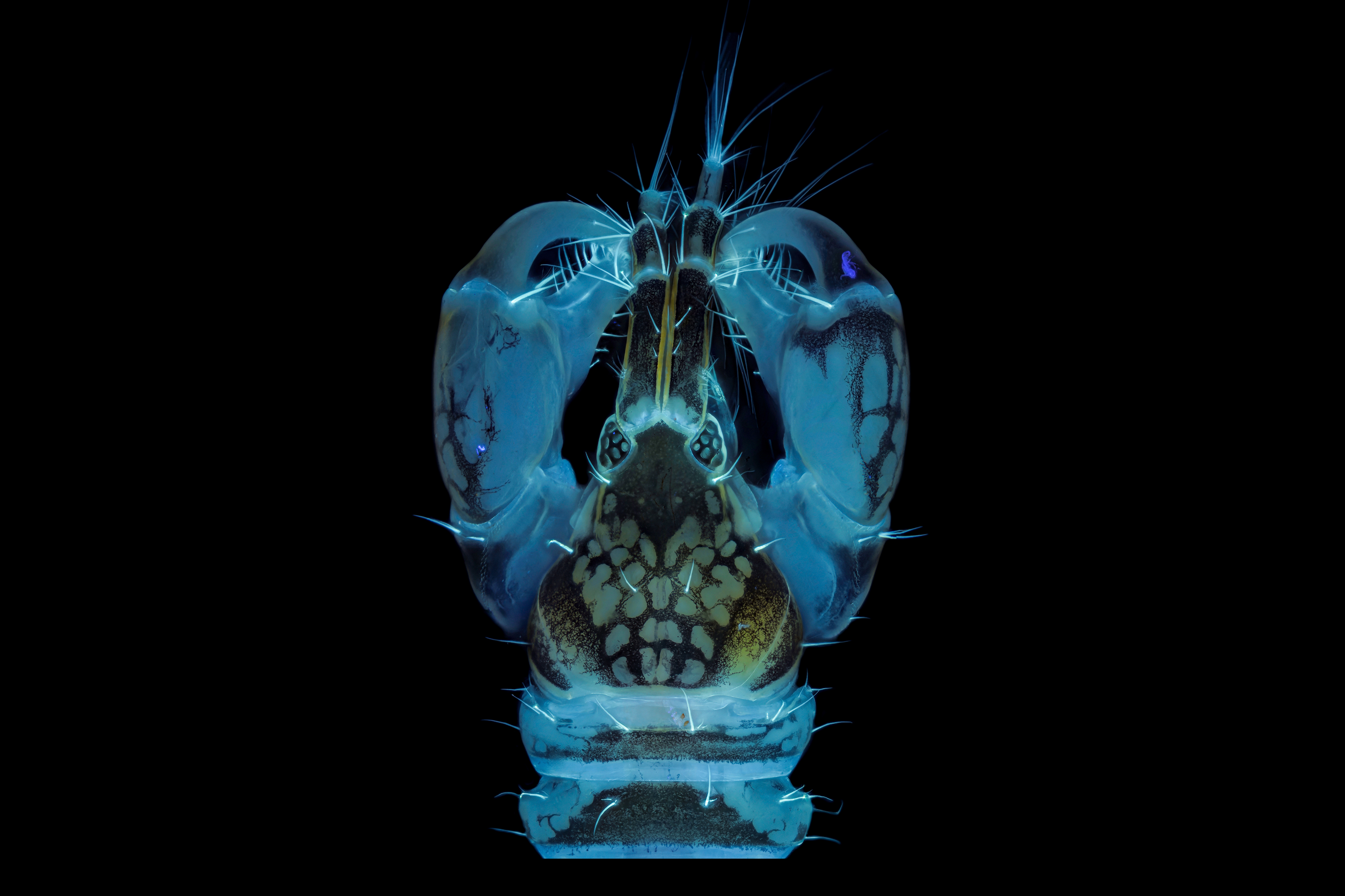
Un crostaceo (Tanaidacea crustacean) mentre nuota, durante la notte, in una colonna d'acqua attratto dalla luce. La chitina emette luce quando viene eccitata dagli UV.

Uno sguardo nelle profondità. Sezioni trasversali dei segmenti apicali dello stelo di una pianta acquatica (Utricularia macrorhiza).

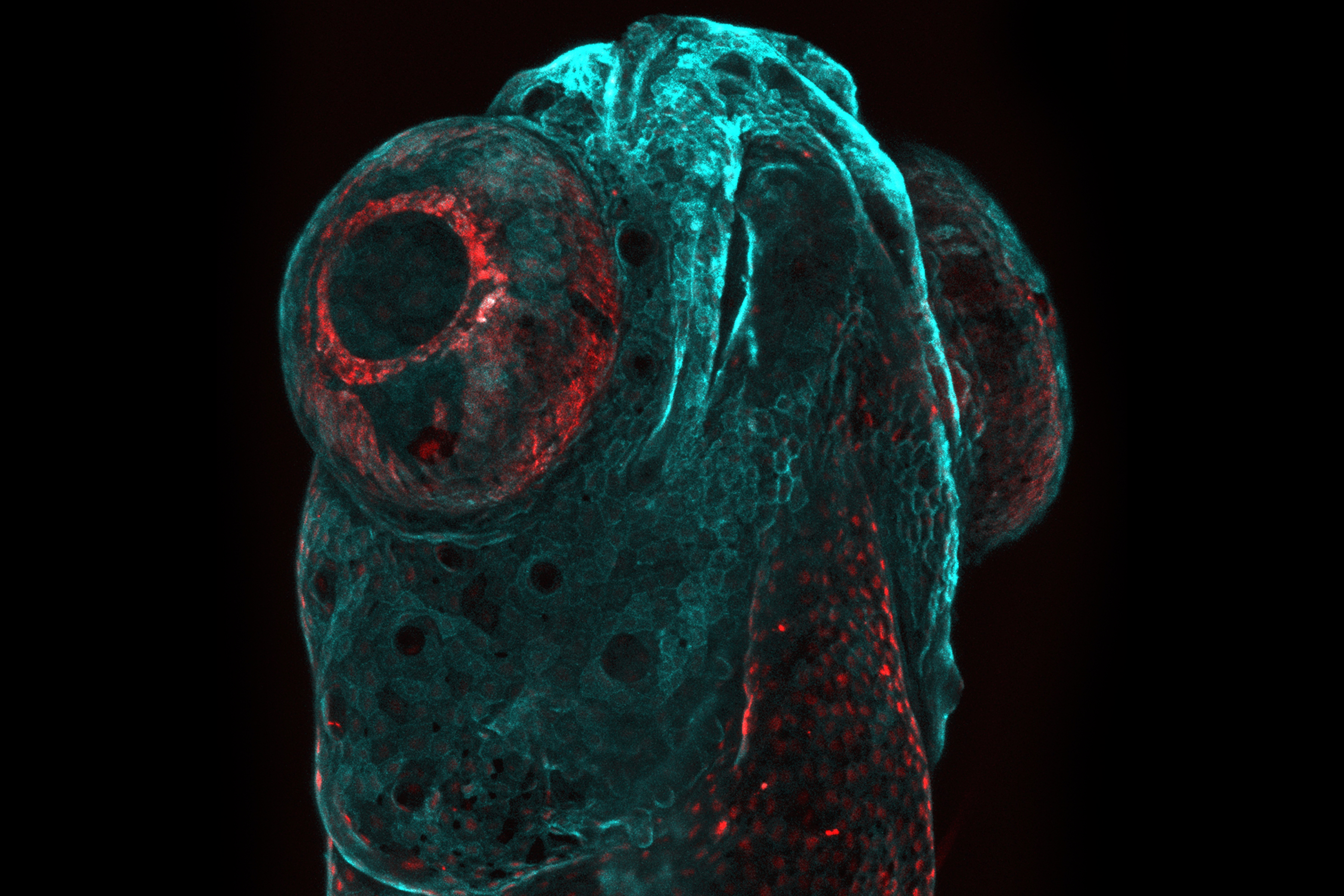
Il sottomarino. L'immagine rappresenta una testa di pesce zebra ricoperto di cellule epiteliali on estrinsecazione della proteina fluorescente palm-mTurquoise e della proteina nucleare mCherry.
Jurors
Americas

Geoff Williams, Manager of the Leduc Bioimaging Facility at Brown University
Geoff Williams is in his 14th year as manager of the Leduc Bioimaging Facility at Brown University. The opportunity to combine visual arts, science, technology, and mastery of a skill clicked with his discovery of microscopy (electron and light) as an undergraduate at Connecticut College. Geoff transitioned from a graduate program at Michigan State University to running the imaging facility at Central Michigan University before arriving at Brown. Over the past 20-plus years, he has been honing his craft as both an electron and light microscopist, paying more attention to the aesthetics of each image collected than is typically required of a purely scientific investigation. Geoff’s work, under the name Nanoscape, provides a tactile and striking view of samples we may or may not encounter in our day-to-day lives.

Harini Sreenivasappa, Manager of the Cell Imaging Center at Drexel University
Harini Sreenivasappa is the manager of Drexel University’s light microscopy core facility, the Cell Imaging Center. She was introduced to microscopy during graduate school at Texas A&M University (TAMU), where she studied the role of microenvironment stimuli on cellular sensing and adapting as it takes place in blood vessel wall remodeling in cardiovascular disease. This led to a PhD in biomedical engineering. She has more than 10 years’ experience working with various microscopy techniques, such as atomic force microscopy (AFM), spinning disk confocal, and total internal reflection fluorescence (TIRF) microscopy. With ASCB’s COMPASS Outreach grant, she created and curated a Traveling Micrographs exhibit showcasing micrographs by TAMU researchers that was free and open to the public. The goal of the series of exhibits was to share research at TAMU with the local community and stimulate interest in imaging science.
EMEA

Rachid Rezgui, Research Instrumentation Scientist, Microscopy, New York University Abu Dhabi
Rachid Rezgui is a microscopist and an active research scientist. Rachid studied physics at the Leibniz University of Hanover in Germany, then completed his PhD in biophysics at the Ecole Polytechnique in France studying DNA-protein interactions at the single molecule level. He joined the microscopy core facility at New York University Abu Dhabi in 2014, and has since worked with all types of microscopes, including two-photon, super-resolution, confocal, fluorescence lifetime, and widefield. He is involved in all aspects of optical imaging, such as sample preparation, training, acquisition, and post-processing, as well as core facility management.

Nicolas Schilling, Microscopy Application Specialist, Center for Microscopy and Image Analysis, University of Zurich
Nicolas Schilling is a microscopy application specialist at the University of Zurich's light and electron microscopy core facility. His journey in optical imaging started during his biomedicine graduate studies at the University of Zurich. After earning his master's degree, he engaged in the university's technology platform program, where he worked on a novel sample preparation method that integrates high-pressure freezing cryofixation with super-resolution light microscopy, enhancing structural preservation. In 2019, he joined the university's microscopy core facility team and has since worked with all types of microscopes, including super-resolution, confocal, widefield, and electron microscopes. His responsibilities are diverse, covering sample preparation, training researchers, data acquisition, and post-processing.
Asia-Pacific

Songhai Shi, Professor and Doctoral Supervisor at the School of Life Sciences, Tsinghua University
Dr. Songhai Shi is a renowned professor and doctoral supervisor at the School of Life Sciences, Tsinghua University in Beijing, China. He has long been dedicated to studying the development, formation, and functioning mechanisms of the brain using methods such as neurobiology, genetics, cell, and developmental biology.

Dr. Wen-Tai Chiu, Professor and Department Chair, Department of Biomedical Engineering, National Cheng Kung University
Dr. Wen-Tai Chiu specializes in optogenetics, calcium signaling, live-cell molecular imaging, and cancer research. He actively serves as a professor at his alma mater, the National Cheng Kung University. His recent projects include investigating the regulation of calcium ions (Ca2+) on focal adhesion dynamics, carcinogenesis, and chemoresistance, as well as exploring advanced bioimaging. His lab has also combined engineering technology to construct an optogenetic platform, which can accurately create different Ca2+ oscillations in time and space scales different from traditional chemical stimulation. As director of the Bioimaging Core Facility at National Cheng Kung University, which is affiliated with the National Core Facility for Biopharmaceuticals of the National Science and Technology Council, Dr. Chiu advocates for the collaborative use of high-end microscopic instruments by internal and external academic and industrial units. He believes that the researchers can learn from each other and practice the ideals of collaboration through the shared platform.
Past Winners


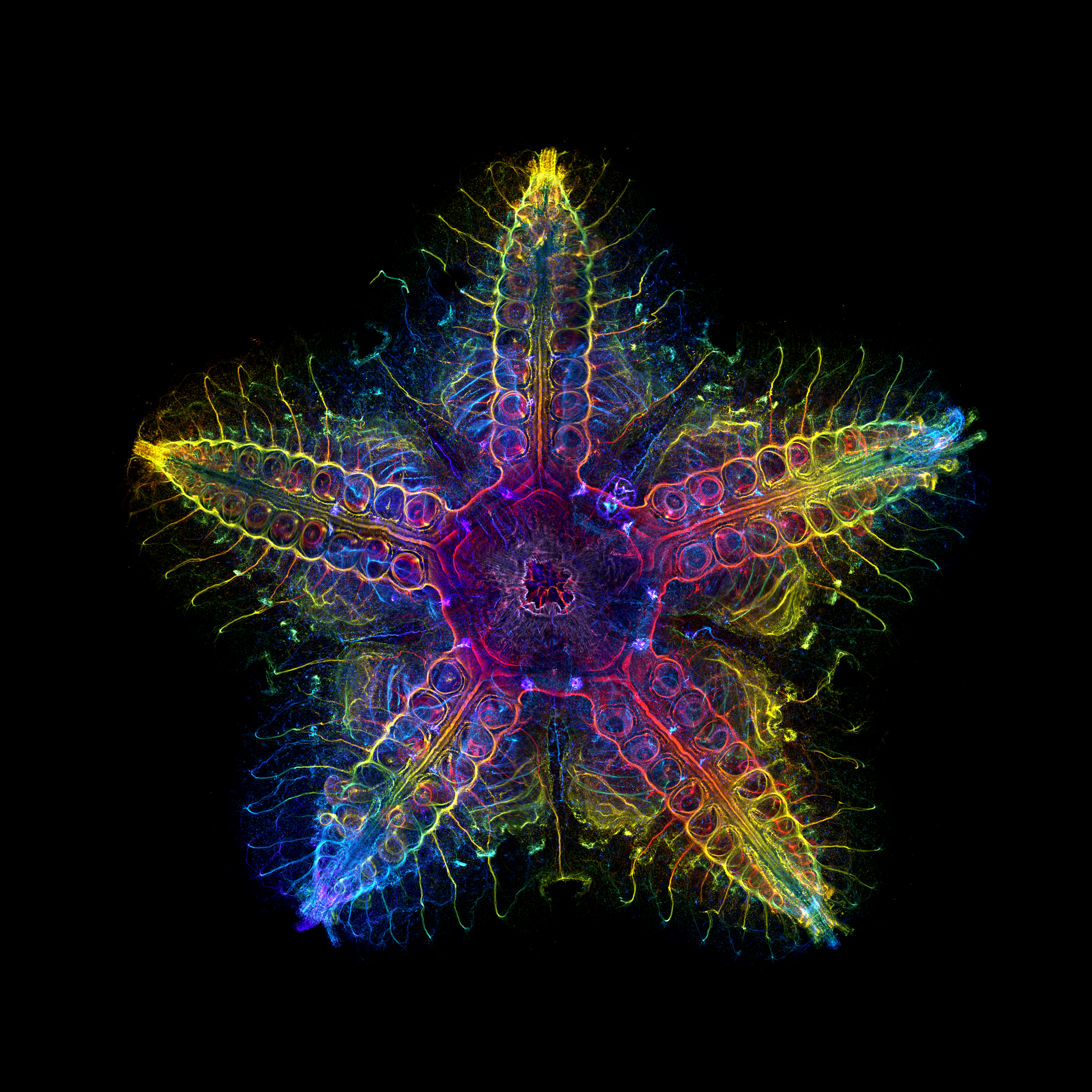
The global winning image was taken by Laurent Formery (USA).
Nervous system of a juvenile sea star (Patiria miniata) about 1 cm wide. Labeled with an antibody against acetylated tubulin after optical clearing, and captured using a color-coded Z-projection.