Where Discovery Begins: MBL and EVIDENT Help Train the Next Generation of Researchers
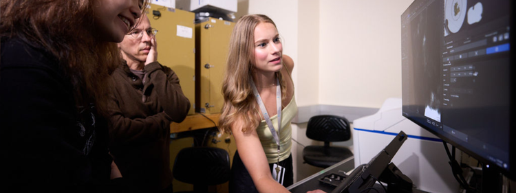
Instructors and students at the MBL observe biology in motion on the Evident APEXVIEW™ APX100 benchtop fluorescence microscope.
Every summer, the Marine Biological Laboratory (MBL) in Woods Hole, Massachusetts, hosts advanced research training courses that give students and early-career researchers hands-on access to the tools, techniques, and scientific environments used in modern biological research.
Through its partnership with the MBL, Evident provides advanced microscopy and imaging systems—including the FLUOVIEW™ FV5000 and IXplore™ IX85 SpinXL confocal systems and the APEXVIEW™ APX100 benchtop fluorescence system—to support live-cell imaging, time-lapse observation, and hands-on experimentation across select courses.
Access to these advanced imaging tools helps students see biological processes more clearly, ask stronger research questions, and build skills they can carry into future scientific work.
Learning Science by Doing Science
The MBL’s advanced research training courses are designed to move beyond lectures and predetermined laboratory exercises. Students work alongside faculty members, teaching assistants, and microscopy specialists on open-ended questions whose answers may not yet be known.
“The MBL advanced research training courses are really incredible vehicles for training the next generation of scientists and for discovery,” said Cliff Brangwynne, co-director of the MBL Physiology course and a faculty member at Princeton University. “It’s not just education. It’s education through doing.”
That distinction shapes the learning environment. Rather than spending a few hours following a standard protocol, participants may work on a project for several weeks, adapting their methods as new observations emerge.
“The discovery process is very hard to teach,” Brangwynne added. “You can’t just hand somebody a protocol or a book and say: this is how you make discoveries.”
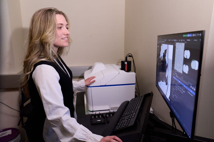
Lauren Todorov, Embryology course participant at the MBL and molecular biology doctoral candidate at Princeton University, uses the APEXVIEW APX100 benchtop fluorescence microscope to perform live-cell imaging.
Michael Hoppa, co-director of the MBL Neurobiology course and a professor of biological sciences at Dartmouth, described the program as an opportunity to understand not only how an instrument works but also how to use it in real research.
“Here, we’re actually trying to get them to have a better understanding of not only how a piece of equipment works, but also to bring projects, do hands-on work, encounter difficulties, and learn how to overcome some of those barriers,” Hoppa noted.
Making Dynamic Biology Visible
Across the advanced MBL courses, microscopy gives participants a way to observe processes that can’t be fully understood through fixed samples or endpoint measurements alone.
Students may watch embryos develop, examine proteins at synaptic interactions, track gene expression, or observe cells as they grow, reorganize, and respond to environmental conditions.
Amy Gladfelter, co-director of the MBL Physiology course and a professor of cell biology and biomedical engineering at Duke University, said the course is built around this direct observation.
“The Physiology course is entirely built around hands-on microscopy,” Gladfelter said. “Nearly every single project and question that gets asked by students in this course involves microscopy.”
Advanced imaging systems enable participants to collect information across both space and time. According to Gladfelter, the resulting combination of spatial detail and temporal resolution can provide a more integrated understanding of cellular behavior.
“What we’re capturing right now with these instruments is a level of resolution, speed, and detail that allows us to really understand the dynamics of cells in real time,” Gladfelter said.
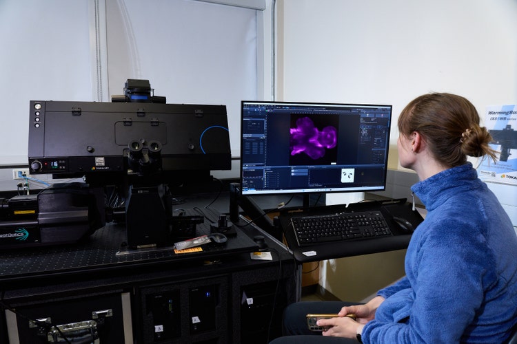
Kensington Hartman, Embryology course participant at the MBL and PhD student in molecular and cell biology at UC Berkeley, performs live-cell imaging on the Evident IXplore™ IX85 SpinXL spinning disk confocal microscope.
In the Physiology course, students have used imaging to localize individual RNA molecules within large fungal cells and quantify gene expression over time and space. They have also observed live slime mold cells as the cells changed their shape, reorganized their networks, and responded to their surroundings.
These experiments are not limited to established laboratory models. Students can also examine organisms collected locally or biological systems that may rarely have been studied with advanced microscopy.
“There are countless examples where the microscopes available here have enabled completely new discoveries,” Gladfelter said. “In some cases, students have visualized things like the cytoskeleton for the very first time in, say, a diatom.”
Choosing the Right Imaging Approach
An important part of microscopy training is learning that no single imaging method is appropriate for every research question.
Live-cell imaging may require low phototoxicity and environmental control. Confocal imaging may be used when optical sectioning or fine structural detail is critical. Time-lapse experiments require a balance of acquisition speed, resolution, sensitivity, and sample health.
“A big part of microscopy, and what we’re teaching in the course, is that it’s a decision-making process,” Gladfelter said. “It’s an optimization problem. What do I care about the most? Do I care the most about seeing this through time? Do I care the most about seeing it through space?”
Having multiple imaging systems available allows students to evaluate these tradeoffs directly. They can compare approaches, adjust acquisition parameters, and select a system based on the specimen and the biological question rather than relying only on the equipment available in their home laboratories.
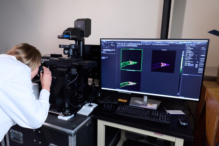
Nadezhda Azbukina, Embryology course participant at the MBL and PhD student in biosystems at ETH Zurich, observes a strain of C. elegans on the Evident FLUOVIEW FV5000 confocal laser scanning microscope.
Athula Wikramanayake, co-director of the MBL Embryology course and professor and chair of biology at the University of Miami, emphasized the growing importance of observing development as it happens.
“There’s more and more interest in doing live imaging to look at developmental processes and the cell biological processes that control embryonic development,” Wikramanayake said. “More and more sensitive ways to look at living embryos during developmental processes are important now, but they’re also going to be the future.”
Broadening Access to Advanced Microscopy and Imaging
Many students arrive at the MBL with substantial expertise in their own research areas but limited access to advanced microscopy and imaging systems. The courses provide time, equipment, and technical support that may not be available at their home institutions.
In the MBL Embryology course, participants work with embryos from approximately 40 animal species and conduct extensive imaging throughout the six-week program.
“They are able to use microscopes here that might not be available in their home labs,” said Tatjana Piotrowski, co-director of the MBL Embryology course and an investigator at the Stowers Institute for Medical Research. “We have microscopy specialists here who help them work with all these microscopes.”
This supported environment helps reduce the hesitation that can accompany unfamiliar equipment. Participants are encouraged to test ideas, learn from unsuccessful experiments, and refine their methods.
“It breaks down the barrier for them to try all these experiments and image as much as they can,” Piotrowski added.
Wikramanayake described this freedom as central to the course experience.
“What we really emphasize in this course is for students to be fearless,” Wikramanayake said. “They can come here, be bold, and do experiments without really having to worry about them failing.”
That freedom can change how participants approach their work after the course. Students return to their institutions with greater confidence in their ability to use imaging, consult core facilities, evaluate technologies, and design experiments outside their previous areas of experience.
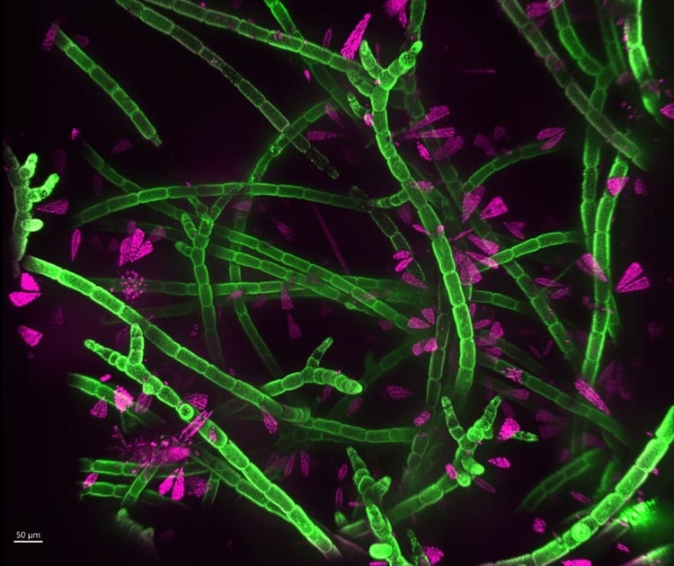
Algae with diatoms, captured on the Evident IXplore IX85 SpinXL spinning disk confocal microscope. Autofluorescence: 488 (algae, green) + 640 (diatoms, magenta). Image courtesy of Madelyn Coble, MBL Embryology course assistant and incoming neuroscience PhD student at UNC–Chapel Hill.
From Training Experiments to Future Research
The work conducted during an MBL course may begin as an exploratory summer project, but its influence can continue well beyond Woods Hole.
Gladfelter refers to the MBL Physiology course as an “idea nursery”—a place where faculty members and students can test experiments that have not yet been explored in depth.
“I would say almost 80 to 90 percent of the experiments that we bring to the course and that are done by students in the course ultimately end up in a publication or some sort of finished product,” Gladfelter said. “Maybe not the microscope image taken in the course, but the experiment is going to be the seed of somebody’s PhD project or a postdoc project.”
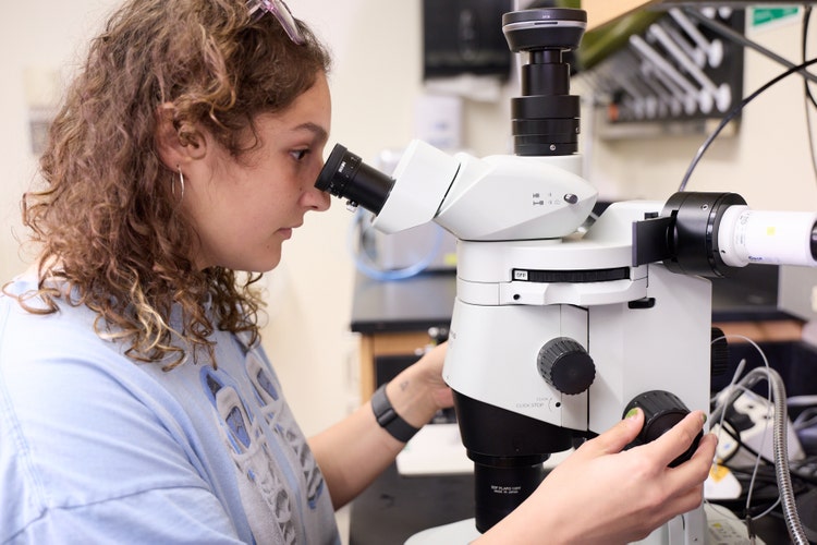
Taylor Pompan, Physiology course teaching assistant at the MBL and third-year graduate student at Duke University, uses the Evident SZX16 stereo microscope.
Many participants also return home prepared to incorporate live-cell imaging, confocal microscopy, quantitative image analysis, or a new model organism into their research.
Scott Dawson, co-director of the MBL Microbial Diversity course and a professor at the University of California, Davis, said the experience can help researchers add imaging to an existing toolkit that may have previously centered on genomics, genetics, or bioinformatics.
“They become more comfortable and realize, ‘I just have to spend some time and look at all the things I can do,’” Dawson said. “They get to add this whole component of imaging to their research toolkit.”
A Partnership Built on Equipment and Expertise
The MBL’s intensive training model creates significant demand for microscope access. Partnership support helps provide the range and quantity of systems needed for students, faculty members, and teaching assistants to conduct experiments throughout the courses.
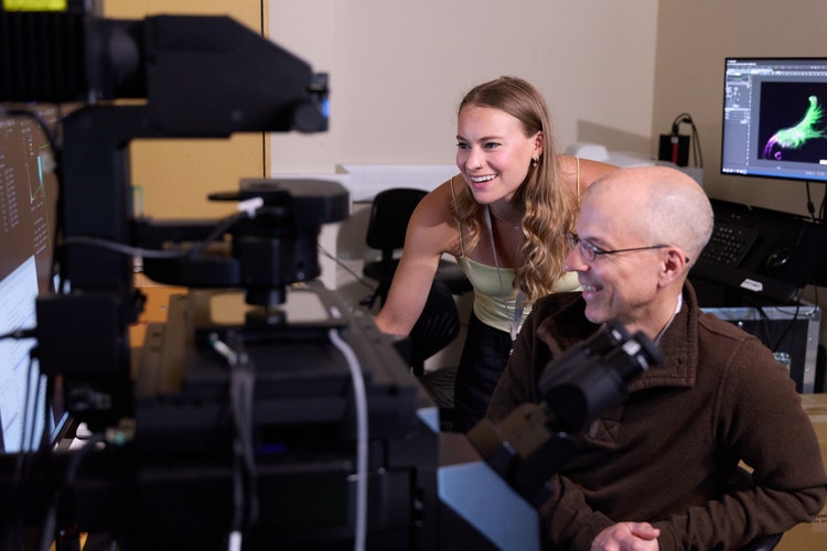
Madelyn Coble, Embryology course assistant at the MBL and incoming neuroscience PhD student at UNC–Chapel Hill, performs imaging on the Evident FLUOVIEW FV5000 confocal laser scanning microscope.
“We could not teach this course without the support of companies like Evident,” Piotrowski said. “There is so much imaging involved with all these embryos that the students are looking at that we need microscopes.”
Hoppa added, “Having access to state-of-the-art equipment, like the FV5000 or Evident objectives for two-photon imaging—where Evident is a leader—means we’re able to really try some of the most challenging experiments.”
The partnership extends beyond providing instruments. Evident specialists can help participants understand system capabilities, optimize acquisition settings, and identify approaches that may not be familiar even to experienced researchers.
“It’s not just the microscopes that come, but also the expertise of the companies that manufacture those microscopes,” said Nipam Patel, director of the MBL. “They can help explain to the students how to optimize their experiments, the details of how their equipment works, and unusual things the equipment can sometimes do that the students—and sometimes the faculty—don’t even know about.”
For instructors, this combination of technology and application support makes it possible to teach microscopy as an active scientific discipline rather than as a set of predefined operating steps.
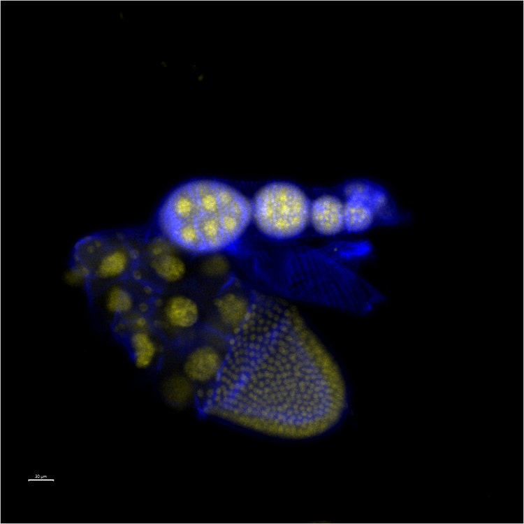
Drosophila ovariole, captured on the Evident FV5000 confocal laser scanning microscope. Antibody stains: 405 (yellow) and 561 (blue). Scale bar: 30 µm. Image courtesy of Madelyn Coble, MBL Embryology course assistant.
For students, it creates an opportunity to live with the systems for several weeks, understand how they perform across different experiments, and make informed decisions about the technologies they may use later in their careers.
“The Marine Biological Laboratory’s advanced research training courses give emerging scientists the opportunity to learn by observing, testing, and discovering for themselves,” said Wes Pringle, Evident CEO. “Through our partnership with the MBL, Evident is proud to help provide the imaging technology and application expertise that enable students and faculty members to explore challenging biological questions. The knowledge and confidence participants develop at Woods Hole can influence the experiments they pursue, the methods they bring back to their institutions, and the laboratories they may lead in the future.”
Inspiring the Next Biological Question
Advanced microscopy does more than produce detailed images. It enables researchers to recognize unexpected patterns, challenge assumptions, and formulate questions that may not have been possible before they saw a process unfold.
“When students come to a course like this and look at their samples and their cells, they realize how much there is to learn,” said Wikramanayake. “They understand that they really are the future and will help us understand development and cell biology going forward.”
For Piotrowski, that future begins with the experience of seeing something that may never have been observed before. “I hope that they really realize how beautiful nature is and how exciting it is to discover new things,” Piotrowski said.
At the MBL, discovery begins with direct observation: a developing embryo, a moving molecule, a changing cell, or an unfamiliar organism brought into focus. Through hands-on research and access to advanced imaging, participants gain more than technical proficiency. They develop the confidence to follow what they see—and to ask where it might lead next.
The APEXVIEW APX100, FLUOVIEW FV5000, and IXplore IX85 SpinXL are for Research Use Only. Not for use in diagnostic procedures.
SZX microscope availability, regulatory status, and intended use vary by country and configuration. Selected models are available for in vitro diagnostic use in certain countries. Products designated for Research Use Only are not for use in diagnostic procedures. Contact your local Evident representative for details.
Related Product
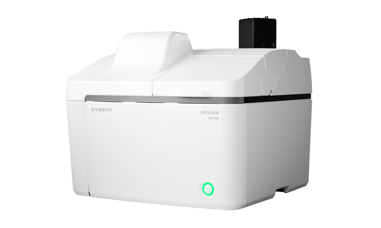
APX100
Benchtop Fluorescence Microscope
The APEXVIEW APX100 benchtop fluorescence microscope makes it fast and simple to acquire expert-quality images. Built with renowned Evident optics, an intuitive user interface, a powerful AI, and a suite of smart features, the APX100 system combines ease of use with high-quality image data to fit your research needs.
- Easy-to-use, all-in-one microscope system
- Publication-quality images in a few clicks
- Fast, efficient data management
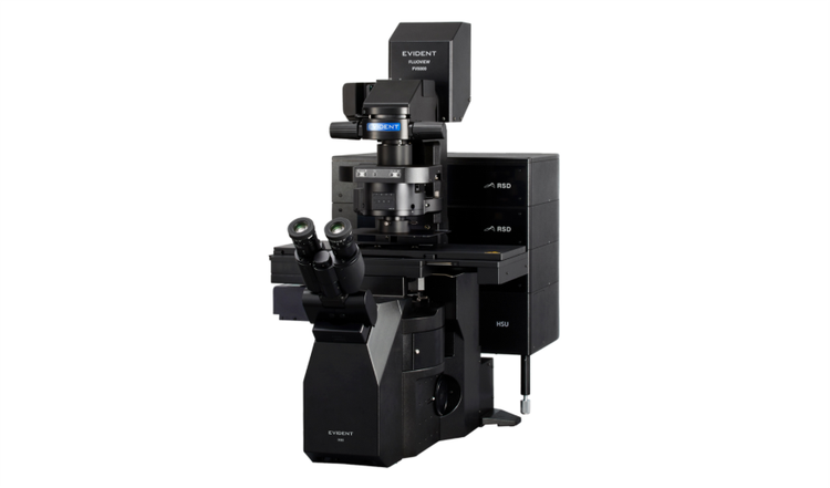
FV5000
Confocal Laser Scanning Microscope
- Extraordinary clarity, speed, and reliability driven by groundbreaking innovations
- SilVIR™ detectors deliver photon-level quantitation, exceptional sensitivity, and ultra-high signal-to-noise
- Unmatched dynamic range captures the full signal spectrum and prevents saturation
- High-speed 2K resonant scanning and high-density 8K galvo scanning in one platform
- FLUOVIEW Smart™ software simplifies operation with intuitive controls and AI-powered automation
- TruResolution™ auto correction collar optimizes focus for over 20 objectives
- Modular design supports up to 10 laser lines and future multiphoton upgrades
- Laser Power Monitor (LPM) ensures stable illumination and reproducible results over time
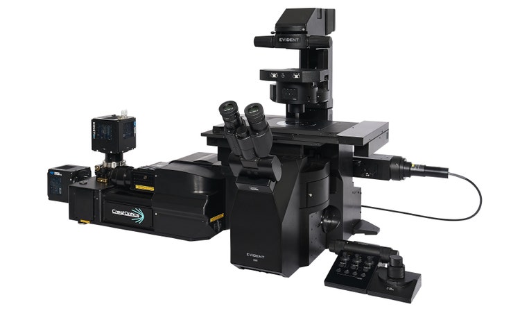
IXplore IX85 SpinXL
Optimized for high-throughput workflows, large datasets, and diverse imaging demands.
- See more and discover faster with an unparalleled 26.5 mm field of view
- Capture more fast cellular dynamics in a single frame with imaging speeds up to 498 fps
- Supports a wide range of applications and users with speed, precision, and flexibility
- Evolves with your research via a wide wavelength range, tailored disk options, and expandable imaging modalities
- Powered by CrestOptics spinning disk technology and enhanced with the IXplore™ IX85 platform